
Image intensifiers are used to convert low-energy x-rays into visible light images. Image intensifiers are several thousand times more sensitive compared to standard 400-speed screen-film combinations, and in practice can produce images using several thousand times less radiation 3,4.
The biggest advantage of image intensifiers in medical imaging is the synergy of high detector efficiency and high conversion efficiency to effectively utilize fluoroscopy while adhering to the radiation protection principle of dose optimization.
Having been used in medical imaging since the 1960's, image intensifiers are far less common in present day due to the introduction of flat-panel detectors (FPD) 7.
On this page:
Components
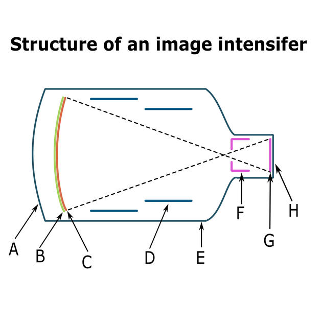
After the x-ray beam emerges from the patient, it enters the image intensifier tube through the input window typically made of low scatter low absorption substances such as titanium or aluminum 1,2 The x-ray beam is then partially absorbed by the fluorescent input screen (entrance phosphor) creating a number of light photons 5.
The light photons strike the photocathode of the input screen and are absorbed by photoelectric interactions, causing it to emit photoelectrons (via the photoelectric effect) 5.
The electrons are accelerated towards the output fluorescent screen by an electric field produced between the photocathode and anode. Focussing and distortion minimization is accomplished by the focussing electrodes 5.
The electrons hit the output phosphor and cause large numbers of light photons to be produced, which subsequently may be captured by various imaging devices 3,4.
Gain and conversion factor
The output of the image intensifier can be evaluated by brightness gain and conversion factor. Brightness gain is the product of minification gain and flux gain (also known as electron gain). Minification gain is the ratio of the input area to output area of phosphor. It makes the image brighter but does not improve the quality or contrast of the image. Meanwhile, flux gain is the number of photons generated at the output phosphor, comparing with photons generated at input phosphor 5.
Brightness gain from minification does not improve the statistical quality of the image because same number of photons are reaching output phosphor whether the input screen is larger or smaller. Thus, the total light output remains the same although minification gain results in increased brightness in the output screen 6.
Conversion factor is defined as output luminance of the image intensifier divided by entrance exposure rate, measuring the efficacy of conversion from x-rays to light. It has units of candela per square meter per milliroentgen per second ([cd/m2]/[mR/sec]) 5.
Artifacts
Vignetting
Vignetting refers to the reduced brightness observed at the peripheries of images captured using II 7. Often, anti-vignetting functions will be applied automatically to correct the image. Factors which contribute to this phenomenon include:
the curved input surface means that the periphery of the input surface is further from the source and thus, due to the inverse square law, will receive a lower dose
less scattered light at the peripheries of the output phosphor
Distortion
Image intensifiers are prone to two main types of distortion:
-
pincushion distortion 7
stretching of the periphery of the image compared to the center
occurs due to the input surface being curved, whilst the output surface is flat
-
S distortion 7
warping of the fluoroscopic image in an 'S' pattern
occurs due to the influence of magnetic fields outside of the II on electrons within the II
the addition of mu-metal to the housing unit of the II attempts to reduce this


 Unable to process the form. Check for errors and try again.