Bell D, Silverstone L, Saber M, Imaging psoas sign (spondylodiscitis). Reference article, Radiopaedia.org (Accessed on 17 Feb 2025) https://doi.org/10.53347/rID-63898
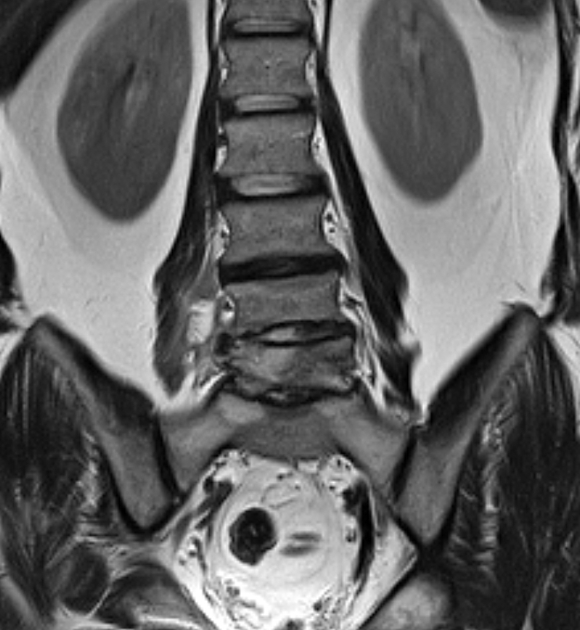
The imaging psoas sign refers to high T2 signal intensity in the psoas muscle which is commonly associated with spondylodiscitis and is a helpful sign in the appropriate clinical context. The psoas muscles have variable origin and attachments 2 to the transverse processes and anteromedial disc and vertebral bodies, commonly T12 to L5.
Other inflammatory and some neoplastic conditions can cause T2 hyperintensity in the psoas muscle, for example myositis and ankylosing spondylitis 1. The original paper 1 demonstrated high sensitivity and specificity in biopsy-confirmed cases of spondylodiscitis however the sign has not been independently validated.
History and etymology
The imaging psoas sign was first described in 2016 by Luke Ledbetter, an American neuroradiologist and his colleagues 1.
1. Ledbetter LN, Salzman KL, Shah LM. Imaging Psoas Sign in Lumbar Spinal Infections: Evaluation of Diagnostic Accuracy and Comparison with Established Imaging Characteristics. (2016) AJNR. American journal of neuroradiology. 37 (4): 736-41. doi:10.3174/ajnr.A4571 - Pubmed
2. Kakarala A, Borthne A, Pierre-Jerome C. MRI of the Psoas Major Muscle: Origin, Attachments, Anatomical Variants and Correlation with Lumbar Disc Extrusion. ARRS 2016. Presentaion-Link


 Unable to process the form. Check for errors and try again.