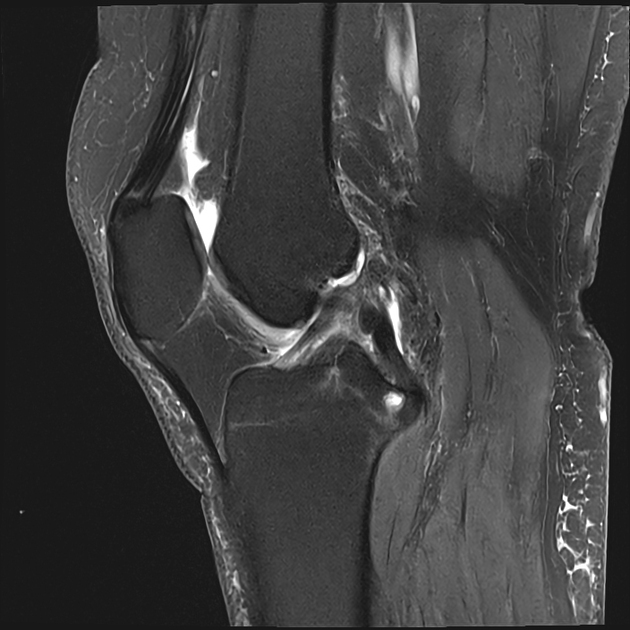
Insertional cysts are usually well-defined, smooth-walled intraosseous cysts found at the insertion sites of tendons and ligaments.
On this page:
Pathology
Etiology
They are thought to be a consequence of bone resorption due to chronic traction and avulsion stresses at the insertional sites of tendons and ligaments 1.
Location
In the knee, they can be found at the insertion sites of the semimembranosus tendon, the cruciate ligaments, or meniscotibial ligaments 1.
Cysts commonly found in the greater and lesser tuberosity of the humeral head at the insertion sites of the supraspinatus and subscapularis tendon could probably fall into the same category 2-4.
Radiographic features
Plain radiograph/CT
Plain radiographs or CT may show small, well-defined cysts.
MRI
MRI will show small, sharply demarcated and well-defined cystic lesions surrounded by a low signal 1.
T1: hypointense
T2: hyperintense
PDFS/T2FS: hyperintense
Surrounding bone marrow edema is very rare 1.
Differential diagnosis
Conditions that can mimic the presentation and/or the appearance of insertional cysts include 1:
-
degenerative subchondral cysts (geodes)
usually located at the opposing sides of the articular surfaces or weight-bearing regions


 Unable to process the form. Check for errors and try again.