Intradiploic epidermoid cysts refer to epidermoid cysts that occur in the diploë of the skull.
On this page:
Clinical presentation
Painless slowly progressive scalp swelling.
Pathology
epidermoid cysts may be congenital (most common, arising from ectodermal inclusion during neural tube closure and subsequently remain within the cranial bones) or acquired (e.g. post-surgical or post-traumatic implantation) 4
intradiploic epidermoids are less frequent than the intradural variety 1
Radiographic features
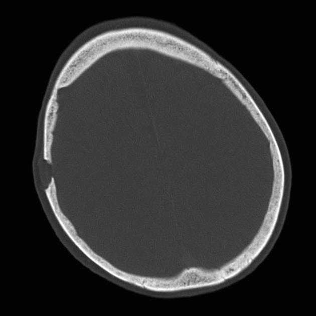
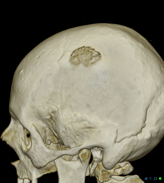
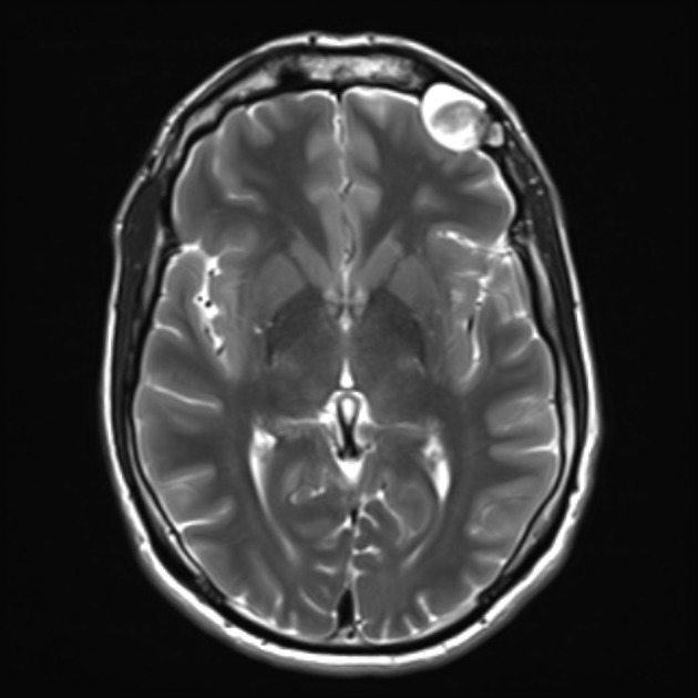
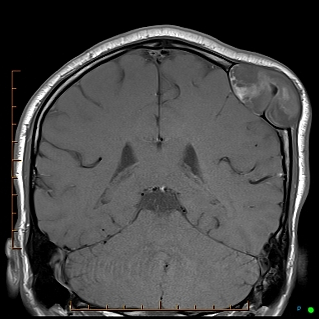
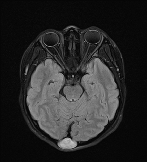
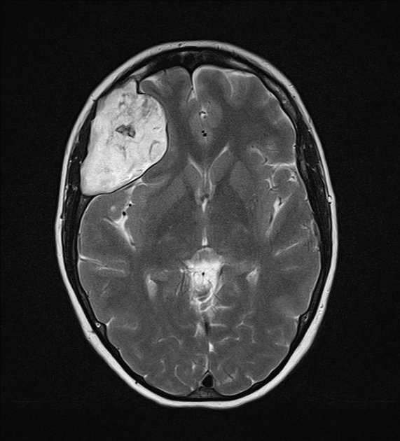
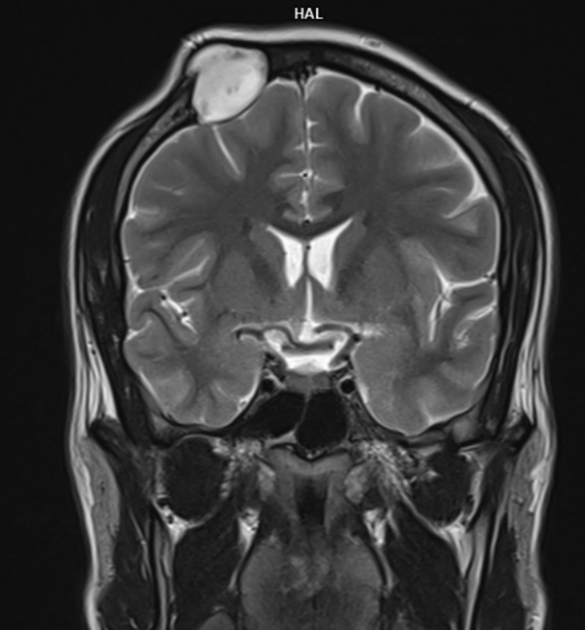
Intradiploic epidermoids occur within the frontal, parietal, occipital and sphenoid bones, as well as the spine 1.
Plain radiograph
rounded or lobulated area of bone destruction, well-delineated sclerotic scalloped margins
CT
non-enhancing hypodense lesion with sharply demarcated bony defects and zones of calcifications
it may alter the outer and/or inner tables of the skull (the inner table more than the outer)
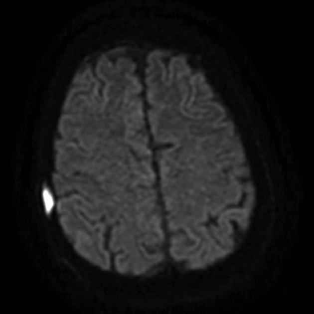
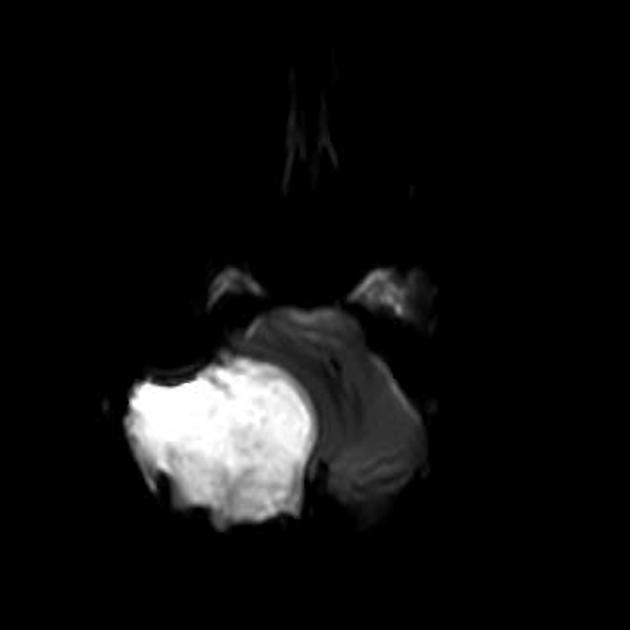
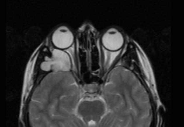
MRI
T1: slightly hyperintense to the CSF
T2: isointense/hyperintense to the CSF
FLAIR: hyperintense to the CSF space
DWI: restricted diffusion with characteristic hyperintensity
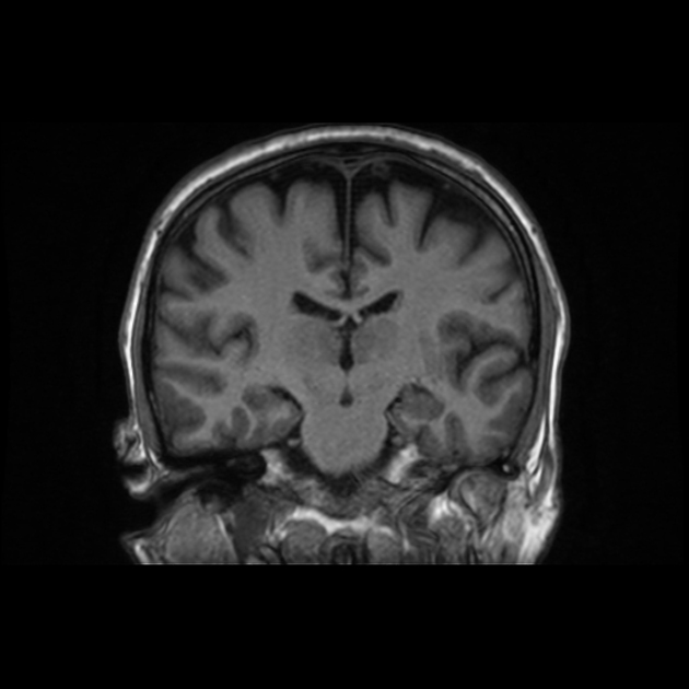
T1C+: none
History and etymology
The first intradiploic epidermoid cyst was reported by J Müller in 1838 5.
The radiological pattern of intradiploic epidermoids was first described by Cushing in 1922.
Differential diagnosis
Consider: