
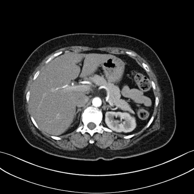
Late arterial phase
Citation, DOI, disclosures and article data
At the time the article was created Yahya Baba had no recorded disclosures.
View Yahya Baba's current disclosuresAt the time the article was last revised Tariq Walizai had no financial relationships to ineligible companies to disclose.
View Tariq Walizai's current disclosures- Late systemic arterial phase
- Arterial systemic phase
- Early venous portal phase
- Hepatic arterial dominant phase
- HAP
- Corticomedullary phase
The late arterial phase, also known as the corticomedullary phase or early venous portal phase, is a contrast-enhanced CT or MRI series, in which there is an optimal enhancement of structures that get their blood supply directly from the arterial system.
The standard characteristics for this phase are:
peak aortic attenuation: 290 - 360 HU 4,5,8
minimal liver enhancement from unenhanced baseline: 20-30 HU 4,6
avid portal vein: density of 145 ± 20 HU 5,8
On this page:
Technique
The acquisition time depends on the intravenous device (central or peripheral), the concentration of the contrast medium, and the injection rate.
-
time from injection through an upper extremity vein: 30-40 seconds 1
30 seconds at an injection rate of 4 ml/s 7
time from bolus tracking: 15-20 seconds
Clinical use
The late arterial phase offers optimal enhancement of the pancreas, spleen, and kidney outer cortex. It allows the detection of:
-
pancreatic lesions
-
liver lesions
-
splenic lesions
See also
References
- 1. Bae KT. Intravenous contrast medium administration and scan timing at CT: considerations and approaches. (2010) Radiology. 256 (1): 32-61. doi:10.1148/radiol.10090908 - Pubmed
- 2. Fleischmann D. Use of high-concentration contrast media in multiple-detector-row CT: principles and rationale. (2003) European radiology. 13 Suppl 5: M14-20. doi:10.1007/s00330-003-2097-z - Pubmed
- 3. Tsuge Y, Kanematsu M, Goshima S, Kondo H, Yokoyama R, Miyoshi T, Onozuka M, Moriyama N, Bae KT. Abdominal vascular and visceral parenchymal contrast enhancement in MDCT: effects of injection duration. (2011) European journal of radiology. 80 (2): 259-64. doi:10.1016/j.ejrad.2010.06.044 - Pubmed
- 4. Sultana S, Awai K, Nakayama Y, Nakaura T, Liu D, Hatemura M, Funama Y, Morishita S, Yamashita Y. Hypervascular hepatocellular carcinomas: bolus tracking with a 40-detector CT scanner to time arterial phase imaging. (2007) Radiology. 243 (1): 140-7. doi:10.1148/radiol.2431060069 - Pubmed
- 5. Yamashita Y, Komohara Y, Takahashi M, Uchida M, Hayabuchi N, Shimizu T, Narabayashi I. Abdominal helical CT: evaluation of optimal doses of intravenous contrast material--a prospective randomized study. (2000) Radiology. 216 (3): 718-23. doi:10.1148/radiology.216.3.r00se26718 - Pubmed
- 6. Murakami T, Kim T, Takamura M, Hori M, Takahashi S, Federle MP, Tsuda K, Osuga K, Kawata S, Nakamura H, Kudo M. Hypervascular hepatocellular carcinoma: detection with double arterial phase multi-detector row helical CT. (2001) Radiology. 218 (3): 763-7. doi:10.1148/radiology.218.3.r01mr39763 - Pubmed
- 7. Monzawa S, Ichikawa T, Nakajima H, Kitanaka Y, Omata K, Araki T. Dynamic CT for detecting small hepatocellular carcinoma: usefulness of delayed phase imaging. (2007) AJR. American journal of roentgenology. 188 (1): 147-53. doi:10.2214/AJR.05.0512 - Pubmed
- 8. Fujigai T, Kumano S, Okada M et al. Optimal Dose of Contrast Medium for Depiction of Hypervascular HCC on Dynamic MDCT. Eur J Radiol. 2012;81(11):2978-83. doi:10.1016/j.ejrad.2012.01.016
Incoming Links
- Zebra sign (disambiguation)
- Hypervascular liver lesions
- Excretory phase
- Hypovascular retroperitoneal lesions
- CT triple-phase liver (protocol)
- Hypervascular retroperitoneal lesions
- Nephrogenic phase
- CT four-phase liver (protocol)
- Hypervascular pancreatic lesions
- Contrast phases
- Early arterial phase
- Hypervascular splenic lesions
- Portal venous phase
Related articles: Computed tomography
- computed tomography in practice
-
computed tomography overview
- iodinated contrast media
- CT IV contrast media administration
-
CT artifacts
- patient-based artifacts
- physics-based artifacts
- hardware-based artifacts
- ring artifact
- tube arcing
- out of field artifact
- air bubble artifact
- helical and multichannel artifacts
- CT technology
-
generations of CT scanners
- helical CT scanning
- step and shoot scanning
- ultra-high-resolution CT (UHRCT)
- CT x-ray tube
- CT fluoroscopy
- cone-beam CT
-
generations of CT scanners
- dual-energy CT
- CT image reconstruction
- CT image quality
- CT dose
-
CT protocols
- composite
- head & neck
- chest
- abdomen and pelvis
- CT abdomen-pelvis (protocol)
- CT abdominal aorta
- CT adrenals (protocol)
- CT cholangiography (protocol)
- CT colonography (protocol)
- CT enteroclysis (protocol)
- CT enterography (protocol)
- CT gastrography (protocol)
- CT kidneys, ureters and bladder (protocol)
- CT urography (protocol)
- CT Renal mass (protocol)
- CT angiography of the splanchnic vessels (protocol)
- CT renal split bolus
- CT pancreas (protocol)
- liver


 Unable to process the form. Check for errors and try again.