Citation, DOI, disclosures and article data
Citation:
Weerakkody Y, Hacking C, Abu Kamesh M, et al. Left atrial appendage thrombus. Reference article, Radiopaedia.org (Accessed on 27 Mar 2025) https://doi.org/10.53347/rID-163481
Disclosures:
At the time the article was last revised Craig Hacking had the following disclosures:
- Philips Australia, Paid speaker at Philips Spectral CT events (ongoing)
These were assessed during peer review and were determined to
not be relevant to the changes that were made.
View Craig Hacking's current disclosures
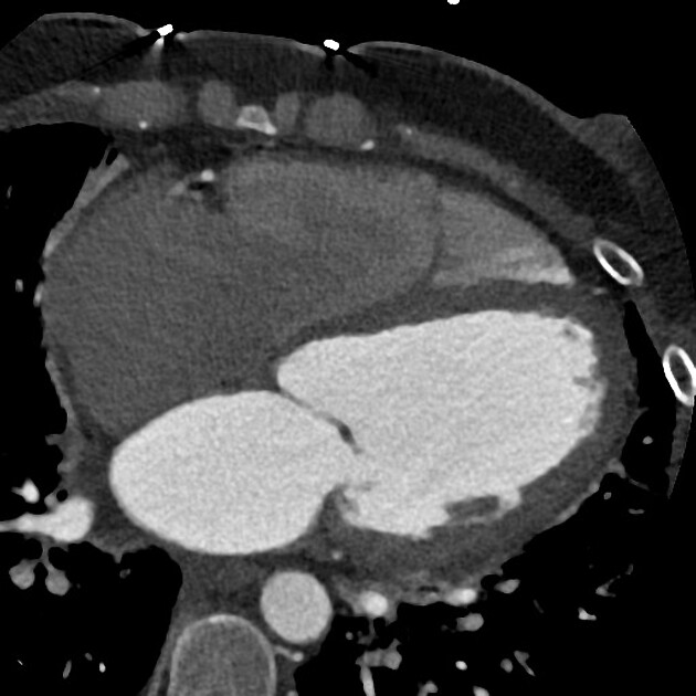
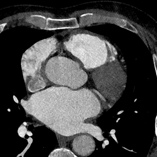
Left atrial appendage thrombus is a site of intra-cardiac thrombus and refers to the presence of thrombus within the left atrial appendage.
The left atrial appendage is considered the main location of thrombus formation, predominantly in patients with non-valvular atrial fibrillation.
Radiographic features
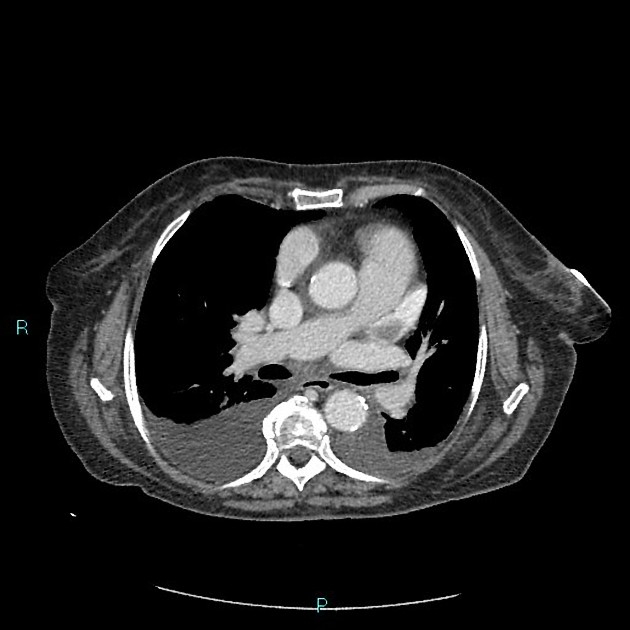
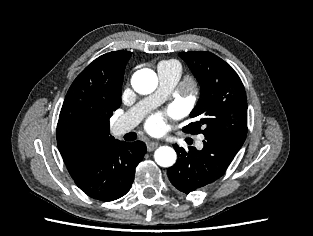
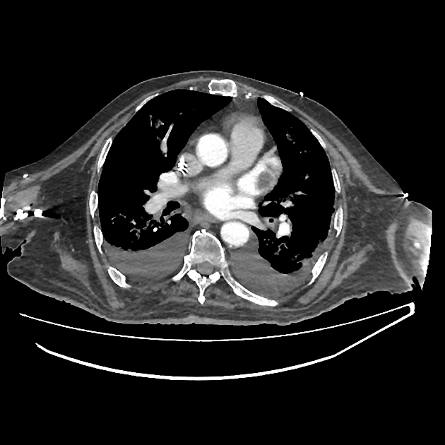
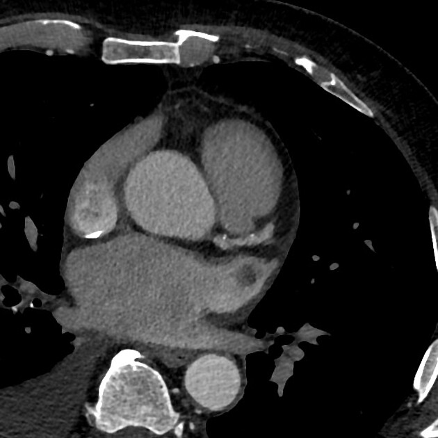
Cardiac CT
Maybe reasonably specific but not as much sensitive 7.
Thrombus detection by cardiac CT relies on filling defects within the left atrial appendage. However, low blood flow velocity may also present as filling defects, and differentiation between thrombi from low blood flow/stasis during early imaging method may be difficult due to the interval between contrast arrival and left atrial appendage image capture being too short. Some authors have suggested a delayed imaging (around 6 minutes) phase to help to differentiate thrombi and low/slow flow 2,5.
Ultrasound
Echocardiography
Trans-esophageal echocardiography (TEE/TOE)
Differential diagnosis
On CT in certain situations consider
-
1. Yuan N, Rader F, Siegel R. Safe to Go with the Flow? Large Left Atrial Appendage Thrombus Despite Robust Appendage Flow Velocities. Eur Heart J Cardiovasc Imaging. 2021;22(6):e76. doi:10.1093/ehjci/jeaa290 - Pubmed
-
2. Yu S, Zhang H, Li H. Cardiac Computed Tomography Versus Transesophageal Echocardiography for the Detection of Left Atrial Appendage Thrombus: A Systemic Review and Meta-Analysis. J Am Heart Assoc. 2021;10(23):e022505. doi:10.1161/JAHA.121.022505 - Pubmed
-
3. Romero J, Cao J, Garcia M, Taub C. Cardiac Imaging for Assessment of Left Atrial Appendage Stasis and Thrombosis. Nat Rev Cardiol. 2014;11(8):470-80. doi:10.1038/nrcardio.2014.77 - Pubmed
-
4. García-Fernández M, Torrecilla E, San Román D et al. Left Atrial Appendage Doppler Flow Patterns: Implications on Thrombus Formation. Am Heart J. 1992;124(4):955-61. doi:10.1016/0002-8703(92)90978-5 - Pubmed
-
5. Spagnolo P, Giglio M, Di Marco D et al. Diagnosis of Left Atrial Appendage Thrombus in Patients with Atrial Fibrillation: Delayed Contrast-Enhanced Cardiac CT. Eur Radiol. 2021;31(3):1236-44. doi:10.1007/s00330-020-07172-2 - Pubmed
-
6. Garcia M. Detection of Left Atrial Appendage Thrombus by Cardiac Computed Tomography: A Word of Caution. JACC Cardiovasc Imaging. 2009;2(1):77-9. doi:10.1016/j.jcmg.2008.10.003 - Pubmed
-
7. Li X, Wang J, Wei Q et al. Diagnostic Value of Delayed Contrast-Enhanced Cardiac Computed Tomography for Detecting Left Atrial Appendage Thrombus in Patients With Atrial Fibrillation. Front Cardiovasc Med. 2022;9:847163. doi:10.3389/fcvm.2022.847163 - Pubmed
Promoted articles (advertising)


 Unable to process the form. Check for errors and try again.