
Lunate morphology
Citation, DOI, disclosures and article data
Citation:
Dixon A, Saber M, Roberts D, et al. Lunate morphology. Reference article, Radiopaedia.org (Accessed on 17 Mar 2025) https://doi.org/10.53347/rID-9811
rID:
9811
Article created:
Disclosures:
At the time the article was created Andrew Dixon had no recorded disclosures.
View Andrew Dixon's current disclosures
Last revised:
Disclosures:
At the time the article was last revised Mohamed Saber had no recorded disclosures.
View Mohamed Saber's current disclosures
Revisions:
14 times, by
11 contributors -
see full revision history and disclosures
Systems:
Sections:
Tags:
Synonyms:
- Lunate types
- Lunate morphological types
Several classification systems exist for the lunate morphology 1, 2.
Classification
The lunate classification proposed by Viegas et al. is arguably the most relevant 3:
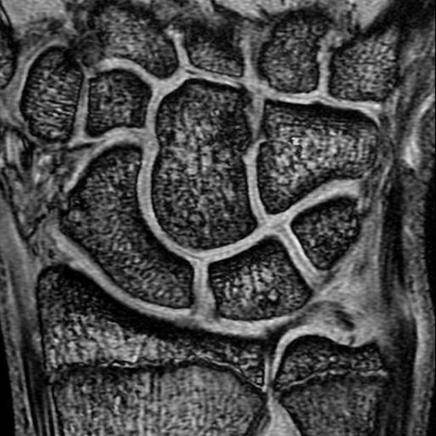
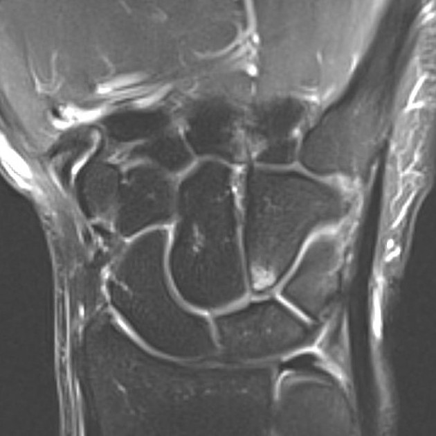
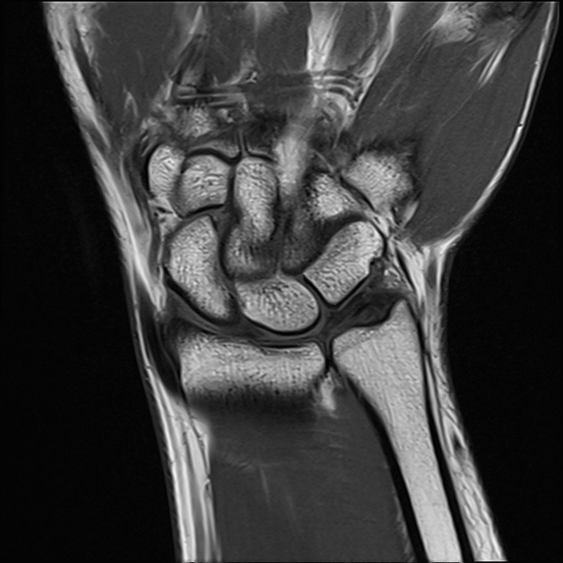
- type I lunate: single distal articular facet for the capitate
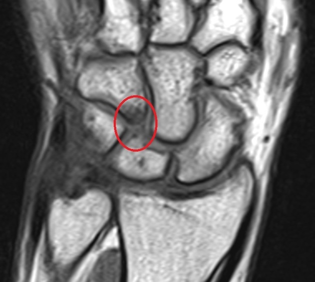
- type II lunate: additional distal articular facet medially for the hamate
There is roughly an even prevalence of the two morphology types.
Related pathology
Type I lunates are associated with a higher prevalence of dorsal intercalated segment instability (DISI) deformity in the setting of scaphoid fracture non-union (~75% vs 20%).
Type II lunates are associated with hamatolunate impingement syndrome leading to a higher prevalence of hamatolunate degenerative arthritis 4.
References
- 1. Taleisnik J. The wrist. Churchill Livingstone. (1985) ISBN:0443081344. Read it at Google Books - Find it at Amazon
- 2. Watson HK, Yasuda M, Guidera PM. Lateral lunate morphology: an x-ray study. J Hand Surg Am. 1996;21 (5): 759-63. doi:10.1016/S0363-5023(96)80188-2 - Pubmed citation
- 3. Viegas SF, Wagner K, Patterson R et-al. Medial (hamate) facet of the lunate. J Hand Surg Am. 1990;15 (4): 564-71. - Pubmed citation
- 4. Cerezal L, Del piñal F, Abascal F et-al. Imaging findings in ulnar-sided wrist impaction syndromes. Radiographics. 22 (1): 105-21. Radiographics (full text) - Pubmed citation
- 5. Malik AM, Schweitzer ME, Culp RW et-al. MR imaging of the type II lunate bone: frequency, extent, and associated findings. AJR. American journal of roentgenology. 173 (2): 335-8. doi:10.2214/ajr.173.2.10430130 - Pubmed
Incoming Links
Articles:
Cases:
- Disrupted Gilula arcs (anatomic variant)
- Bilateral pronator quadratus sign
- Proximal carpal row fracture-dislocation
- Hamatolunate impingement syndrome
- Bifid median nerve and persistant median artery
- Scapholunate fracture with dorsal intercalated segmental instability
- Normal radiographic anatomy of the wrist
- Normal radiographic anatomy of the hand
- Type II lunate with hamatolunate impingement
- Lunate morphology
Related articles: Anatomy: Upper limb
-
skeleton of the upper limb
- clavicle
- scapula
- humerus
- radius
- ulna
- hand
- accessory ossicles of the upper limb
- accessory ossicles of the shoulder
- accessory ossicles of the elbow
-
accessory ossicles of the wrist (mnemonic)
- os centrale carpi
- os epilunate
- os epitriquetrum
- os styloideum
- os hamuli proprium
- lunula
- os triangulare
- trapezium secondarium
- os paratrapezium
- os radiostyloideum (persistent radial styloid)
- joints of the upper limb
-
pectoral girdle
-
shoulder joint
- articulations
- associated structures
- joint capsule
- bursae
- ligaments
- movements
- scapulothoracic joint
-
glenohumeral joint
- arm flexion
- arm extension
- arm abduction
- arm adduction
- arm internal rotation (medial rotation)
- arm external rotation (lateral rotation)
- circumduction
- arterial supply - scapular anastomosis
- ossification centres
-
shoulder joint
-
elbow joint
- proximal radioulnar joint
- ligaments
- associated structures
- movements
- alignment
- arterial supply - elbow anastomosis
- development
-
wrist joint
- articulations
-
ligaments
- intrinsic ligaments
- extrinsic ligaments
- radioscaphoid ligament
- dorsal intercarpal ligament
- dorsal radiotriquetral ligament
- dorsal radioulnar ligament
- volar radioulnar ligament
- radioscaphocapitate ligament
- long radiolunate ligament
- Vickers ligament
- short radiolunate ligament
- ulnolunate ligament
- ulnotriquetral ligament
- ulnocapitate ligament
- ulnar collateral ligament
- associated structures
- extensor retinaculum
- flexor retinaculum
- joint capsule
- movements
- alignment
- ossification centres
-
hand joints
- articulations
- carpometacarpal joint
-
metacarpophalangeal joints
- palmar ligament (plate)
- collateral ligament
-
interphalangeal joints
- palmar ligament (plate)
- collateral ligament
- movements
- ossification centres
- articulations
-
pectoral girdle
- spaces of the upper limb
- muscles of the upper limb
- shoulder girdle
- anterior compartment of the arm
- posterior compartment of the arm
-
anterior compartment of the forearm
- superficial
- intermediate
- deep
-
posterior compartment of the forearm (extensors)
- superficial
- deep
- muscles of the hand
-
accessory muscles
- elbow
- volar wrist midline
- palmaris longus profundus
- aberrant palmaris longus
- volar wrist radial-side
- accessory flexor digitorum superficialis indicis
- flexor indicis profundus
- flexor carpi radialis vel profundus
- accessory head of the flexor pollicis longus (Gantzer muscle, common)
- volar wrist ulnar-side
- dorsal wrist
- blood supply to the upper limb
-
arteries
- subclavian artery (mnemonic)
- axillary artery
- brachial artery (proximal portion)
- ulnar artery
- radial artery
- veins
-
arteries
- innervation of the upper limb
- intercostobrachial nerve
-
brachial plexus (mnemonic)
- branches from the roots
- branches from the trunks
- branches from the cords
- lateral cord
- posterior cord
- medial cord
- terminal branches
- lymphatic drainage of the upper limb


 Unable to process the form. Check for errors and try again.