
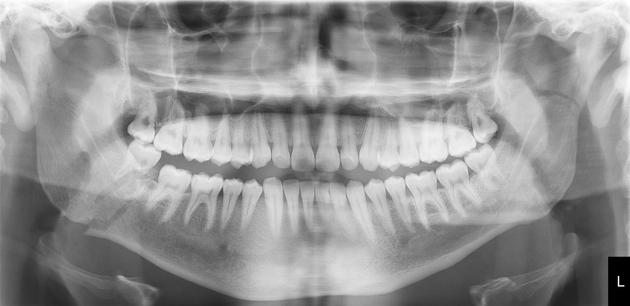
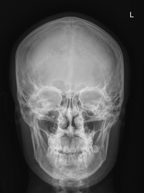
Mandibular fracture
Citation, DOI, disclosures and article data
At the time the article was created Frank Gaillard had no recorded disclosures.
View Frank Gaillard's current disclosuresAt the time the article was last revised Tariq Walizai had no financial relationships to ineligible companies to disclose.
View Tariq Walizai's current disclosures- Mandibular fractures
- Fractures of the mandible
- Fractures involving the mandible
- Mandible fracture
Mandibular fractures are relatively common especially among young men. Although traditionally the mandible and base of skull are thought to form a complete bony ring, interrupted only by the TMJs. This should mean that the mandible should fracture in two places (akin to the bony pelvis) making single fractures uncommon, but this in fact not the case, with ~40% of fractures being unifocal.
On this page:
Epidemiology
After the nasal bones, the mandible is considered the second most common site of facial fractures. Aetiology and demographics will vary significantly depending on the population demographics and with where patients present. In the setting of a trauma centre in New Zealand, 90% of patients are male, with 64% between the ages of 15 and 29 2:
assault: 50%
intimate partner violence
motor vehicle accident (MVA): 10%
fall: 15%
sport: 15%
other: 10%
Clinical presentation
Presenting complaints will include 3:
pain
chin paresthaesia (damage to the mental nerve, a terminal branch of the inferior alveolar nerve)
malocclusion
trismus
dental damage
abnormal mobility
laceration of the skin of the external acoustic meatus
Pathology
Location
angle: 20-33% 1-3
body / horizontal branch: 15-25%
condyle or neck: 15-36% (see: TMJ trauma)
parasymphyseal: 14-15%
ramus: 5%
coronoid process: 1-3%
alveolar ridge: 2%
Unifocal fractures are common, accounting for approximately 40% of all mandibular fractures 1:
multifocal: 60% 1
-
unifocal: 40%
simple: 25%
comminuted: 10%
associated with condylar subluxation: 5%
Subtypes
Treatment and prognosis
Treatment can be conservative or involve formal reduction (which may be open or closed). Closed reduction may be supported with intermaxillary fixation or splints (ORIF).
Complications
permanent malocclusion 3
permanent paraesthesia
References
- 1. Escott EJ, Branstetter BF. Incidence and characterization of unifocal mandible fractures on CT. AJNR Am J Neuroradiol. 2008;29 (5): 890-4. doi:10.3174/ajnr.A0973 - Pubmed citation
- 2. Lee KH. Epidemiology of mandibular fractures in a tertiary trauma centre. Emerg Med J. 2008;25 (9): 565-8. doi:10.1136/emj.2007.055236 - Pubmed citation
- 3. Schwartz DT, Reisdorff EJ. Emergency radiology. McGraw-Hill Professional. (2000) ISBN:0070508275. Read it at Google Books - Find it at Amazon
- 4. Baykul T, Aydın MA, Aksoy MÇ et-al. Unusual unilateral fracture of the condylar and coronoid processes of the mandible. J Clin Imaging Sci. 2014;4 (2): 3. doi:10.4103/2156-7514.141909 - Free text at pubmed - Pubmed citation
- 5. Alessandrino Francesco, Abhishek Keraliya and Jordan Lebovic et al. "Intimate Partner Violence: A Primer for Radiologists to Make the “Invisible” Visible". RadioGraphics 40, no. 7 (2020): 2080-2097. . doi:10.1148/rg.2020200010.
- 6. Dreizin D, Nam A, Tirada N et al. Multidetector CT of Mandibular Fractures, Reductions, and Complications: A Clinically Relevant Primer for the Radiologist. Radiographics. 2016;36(5):1539-64. doi:10.1148/rg.2016150218 - Pubmed
- 7. Nardi C, Vignoli C, Pietragalla M et al. Imaging of Mandibular Fractures: A Pictorial Review. Insights Imaging. 2020;11(1):30. doi:10.1186/s13244-020-0837-0 - Pubmed
Incoming Links
- Mandibular fracture
- Guardsman fracture
- Mandibular fracture
- Subgaleal haematoma
- Mandible fracture
- Mandible fracture
- Facial fracture - Le Fort type III
- Mandibular fracture
- Masticator space blow-out fracture
- Mandibular body and dental fractures
- Mandibular fractures with dental root fracture
- Mandible fractures
- Mandibular ramus fracture
- Mandible fracture (paediatric)
- Mandibular fracture with dental trauma
- Mandibular fractures with dental trauma
- Mandibular fracture
- Mandible fracture
- Mandibular fracture
- Mandibular fracture
Related articles: Fractures
-
fracture
- terminology
- fracture location
- diaphyseal fracture
- metaphyseal fracture
- physeal fracture
- epiphyseal fracture
- fracture types
- avulsion fracture
- articular surface injuries
- complete fracture
- incomplete fracture
- infraction
- compound fracture
- pathological fracture
- stress fracture
- fracture displacement
- fracture location
- fracture healing
- skull fractures
-
facial fractures
- fractures involving a single facial buttress
- alveolar process fractures
- frontal sinus fracture
- isolated zygomatic arch fractures
- mandibular fracture
- nasal bone fracture
- orbital blow-out fracture
- paranasal sinus fractures
- complex fractures
- dental fractures
- fractures involving a single facial buttress
-
spinal fractures
- classification (AO Spine classification systems)
-
cervical spine fracture classification systems
- AO classification of upper cervical injuries
- AO classification of subaxial injuries
- Anderson and D'Alonzo classification (odontoid fracture)
- Roy-Camille classification (odontoid process fracture)
- Gehweiler classifcation (atlas fractures)
- Levine and Edwards classification (hangman fracture)
- Allen and Ferguson classification (subaxial spine injuries)
- subaxial cervical spine injury classification (SLIC)
- thoracolumbar spinal fracture classification systems
- three column concept of spinal fractures (Denis classification)
- classification of sacral fractures
-
cervical spine fracture classification systems
- spinal fractures by region
- spinal fracture types
- classification (AO Spine classification systems)
- rib fractures
- sternal fractures
-
upper limb fractures
- classification
- Rockwood classification (acromioclavicular joint injury)
- AO classification (clavicle fracture)
- Neer classification (clavicle fracture)
- Neer classification (proximal humeral fracture)
- AO classification (proximal humeral fracture)
- AO/OTA classification of distal humeral fractures
- Milch classification (lateral humeral condyle fracture)
- Weiss classification (lateral humeral condyle fracture)
- Bado classification of Monteggia fracture-dislocations (radius-ulna)
- Mason classification (radial head fracture)
- Frykman classification (distal radial fracture)
- Mayo classification (scaphoid fracture)
- Hintermann classification (gamekeeper's thumb)
- Eaton classification (volar plate avulsion injury)
- Keifhaber-Stern classification (volar plate avulsion injury)
- upper limb fractures by region
- shoulder
- clavicular fracture
-
scapular fracture
- acromion fracture
- coracoid process fracture
- glenoid fracture
- humeral head fracture
- proximal humeral fracture
- humeral neck fracture
- arm
- elbow
- forearm
- wrist
-
carpal bones
- scaphoid fracture
- lunate fracture
- capitate fracture
- triquetral fracture
- pisiform fracture
- hamate fracture
- trapezoid fracture
- trapezium fracture
- hand
- shoulder
- classification
- lower limb fractures
- classification by region
- pelvic fractures
- hip fractures
- Pipkin classification (femoral head fracture)
- Garden classification (hip fracture)
- American Academy of Orthopaedic Surgeons classification (periprosthetic hip fracture)
- Cooke and Newman classification (periprosthetic hip fracture)
- Johansson classification (periprosthetic hip fracture)
- Vancouver classification (periprosthetic hip fracture)
- femoral
- knee
- Schatzker classification (tibial plateau fracture)
- AO classification of distal femur fractures
- Meyers and McKeevers classification (anterior cruciate ligament avulsion fracture)
- tibia/fibula
- Watson-Jones classification (tibial tuberosity avulsion fracture)
- ankle
- foot
- Berndt and Harty classification (osteochondral lesions of the talus)
- Sanders CT classification (calcaneal fracture)
- Hawkins classification (talar neck fracture)
- Myerson classification (Lisfranc injury)
- Nunley-Vertullo classification (Lisfranc injury)
- pelvis and lower limb fractures by region
- pelvic fracture
- sacral fracture
- coccygeal fracture
-
hip
- acetabular fracture
- femoral head fracture
-
femoral neck fracture
- subcapital fracture
- transcervical fracture
- basicervical fracture
-
trochanteric fracture
- pertrochanteric fracture
- intertrochanteric fracture
- subtrochanteric fracture
- femur
- mid-shaft fracture
- bisphosphonate-related fracture
- distal femoral fracture
- knee
- avulsion fractures
- Segond fracture
- reverse Segond fracture
- anterior cruciate ligament avulsion fracture
- posterior cruciate ligament avulsion fracture
- arcuate complex avulsion fracture (arcuate sign)
- biceps femoris avulsion fracture
- iliotibial band avulsion fracture
- semimembranosus tendon avulsion fracture
- Stieda fracture (MCL avulsion fracture)
- patellar fracture
- tibial plateau fracture
- avulsion fractures
- leg
- tibial tuberosity avulsion fracture
- tibial shaft fracture
- fibular shaft fracture
- Maisonneuve fracture
- ankle
- foot
- tarsal bones
- metatarsal bones
- phalanges
- classification by region
- terminology


 Unable to process the form. Check for errors and try again.