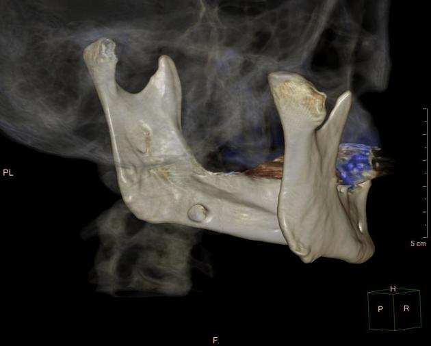
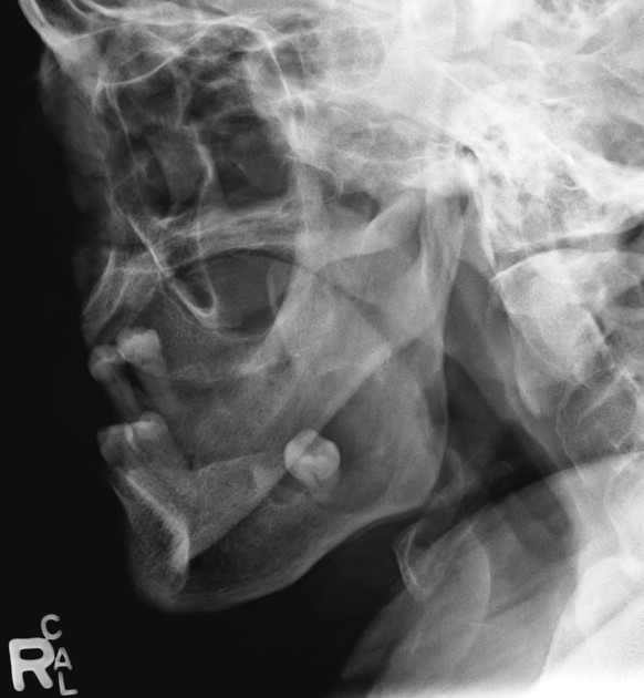
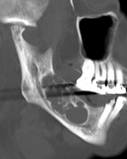
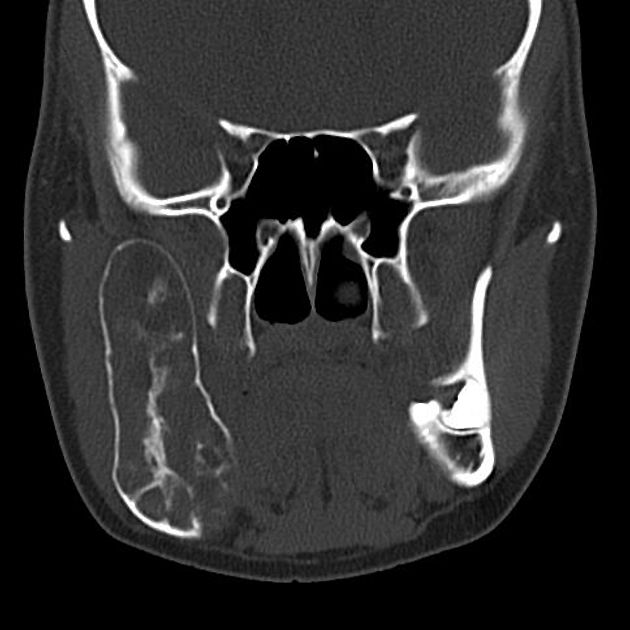
Mandibular lesions are myriad and common. The presence of teeth results in lesions specific to the mandible (and maxilla) and a useful classification that defines them as odontogenic or non-odontogenic. While it may often not be possible to make a diagnosis using imaging alone, this approach to aetiology helps narrow the differential diagnosis.
Pathology
Aetiology
Although a histological approach is probably the most scientifically sound, radiologists are presented with an image. Therefore, it is easier to classify lesions according to their location in the mandible and appearance. For a detailed classification of odontogenic tumours, many more than even the keenest neuro/head and neck radiologists can remember, please refer to the WHO histological classification of odontogenic tumours.
Below, the lesions are divided into cystic and solid. Cystic should not be confused with lytic as solid radiolucent lesions can also appear lytic (see: radiolucent lesions of the jaw).
Cystic lesions
periapical cyst (or radicular cyst): common
dentigerous cyst (or follicular cyst of the mandible): common
odontogenic keratocyst (OKC): uncommon
Stafne cyst (or static bone cavity): uncommon
solitary bone cyst of the mandible (or traumatic or haemorrhagic bone cysts)
aneurysmal bone cyst (ABC): rare in the mandible
Solid lesions
Odontogenic
-
benign
odontoma: most common odontogenic tumour
ameloblastoma: relatively common
odontogenic myxoma (looks just like an ameloblastoma): rare
calcifying epithelial odontogenic tumour (or Pindborg tumour) (looks just like an ameloblastoma): rare
cementoblastoma: rare
-
malignant
odontogenic carcinoma (e.g. ameloblastic carcinoma): rare
odontogenic sarcoma: rare
Non-odontogenic
-
benign
-
malignant


 Unable to process the form. Check for errors and try again.