
Mastoid foramen
Citation, DOI, disclosures and article data
At the time the article was created Prashant Mudgal had no recorded disclosures.
View Prashant Mudgal's current disclosuresAt the time the article was last revised Yuranga Weerakkody had no financial relationships to ineligible companies to disclose.
View Yuranga Weerakkody's current disclosures- Mastoidal emissary foramen
- Mastoid emissary foramina
- Mastoid foramina
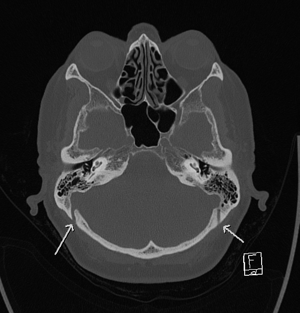
The mastoid foramen is a variably-present foramen as well as being variable in its size, number and position. Most commonly, it is located near the posterior margin of the mastoid process, within the temporo-occipital suture.
Epidemiology
According one publication the prevalence was as follows 3
single foramina on either side : ~ 62.5 %
double foramina on either side: ~ 22.9 %
triple foramina on either side: ~ 6.2 %.
absent: 8.3 % :
It transmits an emissary vein connecting the suboccipital venous plexus to the sigmoid sinus, and the posterior meningeal artery (a small dural branch of the occipital artery).
References
- 1. Imaging of the Head and Neck. Thieme. (2012) ISBN:3131505311. Read it at Google Books - Find it at Amazon
- 2. Wang C, Lockwood J, Iwanaga J, Dumont A, Bui C, Tubbs R. Comprehensive Review of the Mastoid Foramen. Neurosurg Rev. 2020;44(3):1255-8. doi:10.1007/s10143-020-01329-9 - Pubmed
- 3. Murlimanju B, Chettiar G, Prameela M et al. Mastoid Emissary Foramina: An Anatomical Morphological Study with Discussion on Their Evolutionary and Clinical Implications. Anat Cell Biol. 2014;47(3):202-6. doi:10.5115/acb.2014.47.3.202 - Pubmed
- 4. Hampl M, Kachlik D, Kikalova K et al. Mastoid Foramen, Mastoid Emissary Vein and Clinical Implications in Neurosurgery. Acta Neurochir (Wien). 2018;160(7):1473-82. doi:10.1007/s00701-018-3564-2 - Pubmed
Incoming Links
Related articles: Anatomy: Head and neck
- skeleton of the head and neck
-
cranial vault
- scalp (mnemonic)
- fontanelle
-
sutures
- calvarial
- facial
- frontozygomatic suture
- frontomaxillary suture
- frontolacrimal suture
- frontonasal suture
- temporozygomatic suture
- zygomaticomaxillary suture
- parietotemporal suture (parietomastoid suture)
- occipitotemporal suture (occipitomastoid suture)
- sphenofrontal suture
- sphenozygomatic suture
- spheno-occipital suture (not a true suture)
- lacrimomaxillary suture
- nasomaxillary suture
- internasal suture
- basal/internal
- skull landmarks
- frontal bone
- temporal bone
- parietal bone
- occipital bone
- skull base (foramina)
-
facial bones
- midline single bones
- paired bilateral bones
- cervical spine
- hyoid bone
- laryngeal cartilages
-
cranial vault
- muscles of the head and neck
- muscles of the tongue (mnemonic)
- muscles of mastication
-
facial muscles
- epicranius muscle
- circumorbital and palpebral muscles
- nasal muscles
-
buccolabial muscles
- elevators, retractors and evertors of the upper lip
- levator labii superioris alaeque nasalis muscle
- levator labii superioris muscle
- zygomaticus major muscle
- zygomaticus minor muscle
- levator anguli oris muscle
- malaris muscle
- risorius muscle
- depressors, retractors and evertors of the lower lip
- depressor labii inferioris muscle
- depressor anguli oris muscle
- mentalis muscle
- compound sphincter
-
orbicularis oris muscle
- incisivus labii superioris muscle
- incisivus labii inferioris muscle
-
orbicularis oris muscle
- muscle of mastication
- modiolus
- elevators, retractors and evertors of the upper lip
- muscles of the middle ear
- orbital muscles
- muscles of the soft palate
- pharyngeal muscles
- suprahyoid muscles
- infrahyoid muscles
- intrinsic muscles of the larynx
- muscles of the neck
- platysma muscle
- longus colli muscle
- longus capitis muscle
- scalenus anterior muscle
- scalenus medius muscle
- scalenus posterior muscle
- scalenus pleuralis muscle
- sternocleidomastoid muscle
-
suboccipital muscles
- rectus capitis posterior major muscle
- rectus capitis posterior minor muscle
- obliquus capitis superior muscle
- obliquus capitis inferior muscle
- accessory muscles of the neck
- deep cervical fascia
-
deep spaces of the neck
- anterior cervical space
- buccal space
- carotid space
- danger space
- deep cervical fascia
- infratemporal fossa
- masticator space
- parapharyngeal space
- stylomandibular tunnel
- parotid space
- pharyngeal (superficial) mucosal space
- perivertebral space
- posterior cervical space
- pterygopalatine fossa
- retropharyngeal space
- suprasternal space (of Burns)
- visceral space
- surgical triangles of the neck
- orbit
- ear
- paranasal sinuses
- upper respiratory tract
- viscera of the neck
- blood supply of the head and neck
-
arterial supply
-
common carotid artery
- carotid body
- carotid bifurcation
- subclavian artery
- variants
-
common carotid artery
- venous drainage
-
arterial supply
- innervation of the head and neck
-
cranial nerves
- olfactory nerve (CN I)
- optic nerve (CN II)
- oculomotor nerve (CN III)
- trochlear nerve (CN IV)
-
trigeminal nerve (CN V) (mnemonic)
- trigeminal ganglion
- ophthalmic division
- maxillary division
- mandibular division
- abducens nerve (CN VI)
- facial nerve (CN VII)
-
vestibulocochlear nerve (CN VIII)
- vestibular ganglion (Scarpa's ganglion)
- glossopharyngeal nerve (CN IX)
- vagus nerve (CN X)
- (spinal) accessory nerve (CN XI)
- hypoglossal nerve (CN XII)
- parasympathetic ganglia of the head and neck
- cervical sympathetic ganglia
- greater occipital nerve
- third occipital nerve
-
cervical plexus
- muscular branches
- longus capitis
- longus colli
- scalenes
- geniohyoid
- thyrohyoid
-
ansa cervicalis
- omohyoid (superior and inferior bellies separately)
- sternothyroid
- sternohyoid
- phrenic nerve
- contribution to the accessory nerve (CN XI)
- cutaneous branches
- muscular branches
- brachial plexus
- pharyngeal plexus
-
cranial nerves
- lymphatic drainage of the head and neck
- embryological development of the head and neck


 Unable to process the form. Check for errors and try again.