
This is a basic article for medical students and other non-radiologists
MRI brain is a specialist investigation that is used for the assessment of a number of neurological conditions. It is the main method to investigate conditions such as multiple sclerosis and headaches, and used to characterize strokes and space-occupying lesions.
On this page:
Reference article
This is a summary article; we do not have a more in-depth reference article.
Summary
-
indications
confirmation of stroke
assessment of intracranial tumor
chronic headache
seizure disorder
-
important pathology
-
benefits
multiplanar assessment of the brain
exceptionally detailed images of the brain
different sequences allow assessment of different pathology
no ionizing radiation (especially important in children)
-
limitations
much longer investigation (20-40 minutes)
less available (longer waiting list)
patients may be claustrophobic
-
contraindicated in patients with some metallic implants
most pacemakers are not MRI-compatible
-
procedure
patient positioned on the MRI couch
head coil positioned over their head
patient moved into the center of the magnet
sequences acquired
-
similar tests
-
first-line investigation in most acute situations
-
CT head with contrast
initial assessment of intracranial lesions
-
About MRI
Different pulses and different signals provide a variety of sequences and images that we use. Unlike CT where we describe "density", images are described by signal intensity ("hyper-" bright, "hypo-" dark).
-
T1
provides the most anatomically-relevant images
fluid (in CSF and orbits) is dark
grey matter is darker than the white matter
-
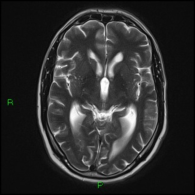
T2
standard sequence
fluid is bright
white matter is darker than grey
-
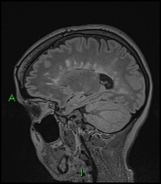
FLAIR (fluid attenuation inversion recovery)
commonly used sequence
similar to T2, but the fluid is darker or "suppressed"
useful for areas of edema or inflammation
used to identify plaques in multiple sclerosis (especially periventricular)
-
DWI and ADC (diffusion-weighted imaging and apparent diffusion coefficient)
these "blocky" images show how easily water moves around
restricted diffusion occurs in stroke, abscesses and cellular tumors


 Unable to process the form. Check for errors and try again.