Optic neuritis
Updates to Article Attributes
Optic neuritis merely denotes inflammation of the optic nerve, and is one of the more common causes of optic neuropathy. It can be thought of as broadly divided into infectious and noninfectious causes, although the latter is far more frequent. On imaging, optic neuritis is most most easily identified as a unilateral optic nerve swelling, with high T2 signal and contrast enhancement.
Epidemiology
The demographics of people affected will conform to those of the underlying conditions (see Pathology).
As typical optic neuritis is seen in the setting of multiple sclerosis (MS), most patients tend to be young adults, with a predominance of women of 3:1.
The incidence is highest in populations living at higher northern latitudes (e.g. Scandinavia, United Kingdom, Canada), again, following the epidemiology of MS 3.
Clinical presentation
Typical optic neuritis (that seen in the setting of demyelination) causes pain in the orbit (90%), often worse with eye movement, and is associated with visual loss, which reaches a nadir within a few days of symptom onset 1,4. The degree of visual loss is variable, ranging from minimal if any visual loss to complete absence of light perception. Additionallydyschromatopsia, photopsia and visual field defects may also occur 1. Fundoscopy typically demonstrates diffuse disc swelling 1.
Pathology
In the majority of cases, typical optic neuritis (as is encountered inmultiple sclerosis) is unilateral1. In contrast, in the setting ofneuromyelitis optica (NMO), involvement is usually bilateral.
Aetiology
Optic neuritis can arise in the setting of many infective and non-infective conditions:
- non-infective
- multiple sclerosis (most common)
- neuromyelitis optica (NMO)
- sarcoidosis
- acute disseminated encephalomyelitis (ADEM)
- systemic lupus erythematosus (SLE)
- toxins
- radiation-induced
- infective
- Lyme disease
- toxoplasmosis
- human immunodeficiency virus (HIV)
- systemic viral diseases asvaricella orherpes
Radiographic features
MRI is the modality of choice for visualising the optic nerve. Functional MRI or multifocal multifocal visual evoked potentials have also been shown to allow early diagnosis 1.
MRI
Typically findings are most easily identified in the retrobulbar intra-orbital segment of the optic nerve, which appears swollen, with high T2 signal. High T2 signal persists and may be permanent; chronically the nerve will appear atrophied rather than swollen.
Contrast enhancement of the nerve, best seen with fat-suppressed T1 coronal images, is seen in >90% of patients if scanned within 20 days of visual loss 2.
Treatment and prognosis
Typical optic neuritis is self-limiting, and recovery of vision usually begins within a few weeks of symptom onset 1.
Although there is considerable evidence that corticosteroid therapy does not alter visual outcome at six months, it does appear to hasten recovery and some trials have shown persistent improvement of contrast contrast sensitivity, visual fields, and colour vision 1.
As such, if therapy is prescribed, it is usually 3 or more days of high dose steroids, started as early after symptom onset as possible 1.
-<p><strong>Optic neuritis</strong> merely denotes inflammation of the <a href="/articles/optic-nerve">optic nerve</a>, and is one of the more common causes of <a href="/articles/optic-neuropathy">optic neuropathy</a>. It can be thought of as broadly divided into infectious and noninfectious causes, although the latter is far more frequent. On imaging, optic neuritis is most easily identified as a unilateral optic nerve swelling, with high T2 signal and contrast enhancement.</p><h4>Epidemiology</h4><p>The demographics of people affected will conform to those of the underlying conditions (see Pathology). </p><p>As typical optic neuritis is seen in the setting of multiple sclerosis (MS), most patients tend to be young adults, with a predominance of women of 3:1. </p><p>The incidence is highest in populations living at higher northern latitudes (e.g. Scandinavia, United Kingdom, Canada), again, following the epidemiology of MS <sup>3</sup>. </p><h4>Clinical presentation</h4><p>Typical optic neuritis (that seen in the setting of demyelination) causes pain in the orbit (90%), often worse with eye movement, and is associated with visual loss, which reaches a nadir within a few days of symptom onset <sup>1,4</sup>. The degree of visual loss is variable, ranging from minimal if any visual loss to complete absence of light perception. Additionally <a href="/articles/dyschromatopsia">dyschromatopsia</a>, <a href="/articles/photopsia">photopsia</a> and visual field defects may also occur <sup>1</sup>. Fundoscopy typically demonstrates diffuse disc swelling <sup>1</sup>. </p><h4>Pathology</h4><p>In the majority of cases, typical optic neuritis (as is encountered in <a href="/articles/multiple-sclerosis">multiple sclerosis</a>) is unilateral <sup>1</sup>. In contrast, in the setting of <a href="/articles/neuromyelitis-optica">neuromyelitis optica (NMO)</a>, involvement is usually bilateral. </p><h5>Aetiology</h5><p>Optic neuritis can arise in the setting of many infective and non-infective conditions:</p><ul>- +<p><strong>Optic neuritis</strong> merely denotes inflammation of the <a href="/articles/optic-nerve">optic nerve</a>, and is one of the more common causes of <a href="/articles/optic-neuropathy">optic neuropathy</a>. It can be thought of as broadly divided into infectious and noninfectious causes, although the latter is far more frequent. On imaging, optic neuritis is most easily identified as a unilateral optic nerve swelling, with high T2 signal and contrast enhancement.</p><h4>Epidemiology</h4><p>The demographics of people affected will conform to those of the underlying conditions (see Pathology). </p><p>As typical optic neuritis is seen in the setting of multiple sclerosis (MS), most patients tend to be young adults, with a predominance of women of 3:1. </p><p>The incidence is highest in populations living at higher northern latitudes (e.g. Scandinavia, United Kingdom, Canada), again, following the epidemiology of MS <sup>3</sup>. </p><h4>Clinical presentation</h4><p>Typical optic neuritis (that seen in the setting of demyelination) causes pain in the orbit (90%), often worse with eye movement, and is associated with visual loss, which reaches a nadir within a few days of symptom onset <sup>1,4</sup>. The degree of visual loss is variable, ranging from minimal if any visual loss to complete absence of light perception. Additionally <a href="/articles/dyschromatopsia">dyschromatopsia</a>, <a href="/articles/photopsia">photopsia</a> and visual field defects may also occur <sup>1</sup>. Fundoscopy typically demonstrates diffuse disc swelling <sup>1</sup>. </p><h4>Pathology</h4><p>In the majority of cases, typical optic neuritis (as is encountered in <a href="/articles/multiple-sclerosis">multiple sclerosis</a>) is unilateral <sup>1</sup>. In contrast, in the setting of <a href="/articles/neuromyelitis-optica">neuromyelitis optica (NMO)</a>, involvement is usually bilateral. </p><h5>Aetiology</h5><p>Optic neuritis can arise in the setting of many infective and non-infective conditions:</p><ul>
-<li><a href="/articles/missing">Lyme disease</a></li>- +<li><a href="/articles/peroneus-brevis-1">Lyme disease</a></li>
-<li>systemic viral diseases as <a href="/articles/varicella-pneumonia">varicella</a> or <a href="/articles/herpes-simplex-encephalitis">herpes </a> </li>- +<li>systemic viral diseases as <a href="/articles/varicella-pneumonia">varicella</a> or <a href="/articles/herpes-simplex-encephalitis">herpes </a> </li>
-</ul><h4>Radiographic features</h4><p>MRI is the modality of choice for visualising the optic nerve. Functional MRI or multifocal visual evoked potentials have also been shown to allow early diagnosis <sup>1</sup>. </p><h5>MRI</h5><p>Typically findings are most easily identified in the retrobulbar intra-orbital segment of the optic nerve, which appears swollen, with high T2 signal. High T2 signal persists and may be permanent; chronically the nerve will appear atrophied rather than swollen. </p><p>Contrast enhancement of the nerve, best seen with fat-suppressed T1 coronal images, is seen in >90% of patients if scanned within 20 days of visual loss <sup>2</sup>. </p><h4>Treatment and prognosis</h4><p>Typical optic neuritis is self-limiting, and recovery of vision usually begins within a few weeks of symptom onset <sup>1</sup>. </p><p>Although there is considerable evidence that corticosteroid therapy does not alter visual outcome at six months, it does appear to hasten recovery and some trials have shown persistent improvement of contrast sensitivity, visual fields, and colour vision <sup>1</sup>. </p><p>As such, if therapy is prescribed, it is usually 3 or more days of high dose steroids, started as early after symptom onset as possible <sup>1</sup>. </p>- +</ul><h4>Radiographic features</h4><p>MRI is the modality of choice for visualising the optic nerve. Functional MRI or multifocal visual evoked potentials have also been shown to allow early diagnosis <sup>1</sup>. </p><h5>MRI</h5><p>Typically findings are most easily identified in the retrobulbar intra-orbital segment of the optic nerve, which appears swollen, with high T2 signal. High T2 signal persists and may be permanent; chronically the nerve will appear atrophied rather than swollen. </p><p>Contrast enhancement of the nerve, best seen with fat-suppressed T1 coronal images, is seen in >90% of patients if scanned within 20 days of visual loss <sup>2</sup>. </p><h4>Treatment and prognosis</h4><p>Typical optic neuritis is self-limiting, and recovery of vision usually begins within a few weeks of symptom onset <sup>1</sup>. </p><p>Although there is considerable evidence that corticosteroid therapy does not alter visual outcome at six months, it does appear to hasten recovery and some trials have shown persistent improvement of contrast sensitivity, visual fields, and colour vision <sup>1</sup>. </p><p>As such, if therapy is prescribed, it is usually 3 or more days of high dose steroids, started as early after symptom onset as possible <sup>1</sup>. </p>
Image 2 MRI (T2 orbit) ( update )
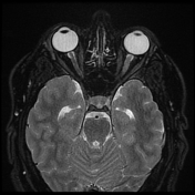
Image 4 MRI (T2) ( create )

Image 5 MRI (T2) ( create )
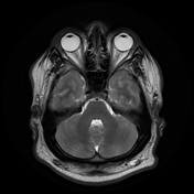
Image 6 MRI (T1 C+ fat sat) ( create )
