
The optic nerve is the second (CN II) cranial nerve (TA: nervus opticus or nervus cranialis II). It is a purely sensory nerve that conveys visual information from the eye to the brain.
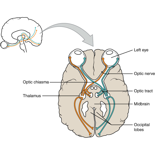
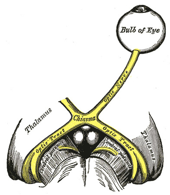
The nerve arises from the back of the globe exiting the orbit via the optic canal. It joins the contralateral optic nerve at the optic chiasm where medial fibers decussate before continuing as the optic tracts.
The cells of origin consist of the ganglion cells of the retina with the main central connections consisting of the lateral geniculate nucleus of the thalamus, and the pretectal area of the midbrain.
On this page:
Gross anatomy
Similar to the olfactory nerve (CN I), the optic nerve is really an extension of the central nervous system. It is not surrounded by Schwann cells with the first sensory bipolar cell body located peripherally in the retina. Their central processes synapse on ganglion cells on the vitreous surface of the retina and their central processes pass via the optic disc out of the globe and form the optic nerve proper. The optic nerve is traditionally divided into four segments: intraocular, intraorbital, intracanalicular, and intracranial.
Intraocular segment
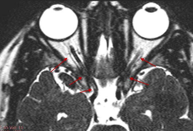
Formed from nerve fibers of the retina and emerges through an opening in the sclera known as the lamina cribrosa. It is approximately 1 mm in length 8.
Intraorbital segment
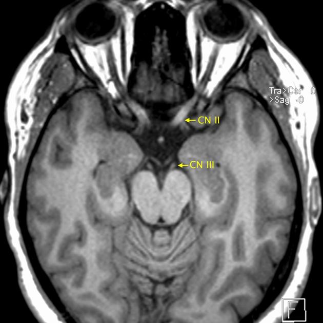
Passes posteriorly and centrally within the orbit and is surrounded by dural lining and CSF; hence it directly communicates with the subarachnoid space and therefore allows transmission of increased pressure from hydrocephalus to manifest as papilledema; additionally, the dural covering can develop a meningioma. It is approximately 25 mm in length 8.
Intracanalicular segment
Where the optic nerve exits through the tendinous ring and optic canal superior to the ophthalmic artery. It is approximately 9 mm in length 8.
Intracranial segment
Also known as the cisternal or prechiasmatic segment, enters the middle cranial fossa and passes within the suprasellar cistern with the anterior cerebral artery at its superolateral aspect joining the contralateral optic nerve at the optic chiasm. It is approximately 16 mm in length 8.
At the optic chiasm, the nasal fibers of each optic nerve (fibers carrying light impulses from the nasal side of the retina) decussate while the temporal fibers do not (partial decussation). From the optic chiasm arise two optic tracts, each one containing nasal fibers of the contralateral optic nerve and temporal fibers from the ipsilateral optic nerve. The optic tract courses around the cerebral peduncle to relay in the lateral geniculate body of the thalamus.
Arterial supply
Intraocular, intraorbital, and intracanalicular segments are supplied by the ophthalmic artery and its branch, the central retinal artery.
Small branches of the anterior cerebral artery (ACA) and the superior hypophyseal artery supply the intracranial segment of the optic nerves and optic chiasm.
The optic tracts are supplied by small branches of the anterior choroidal and posterior communicating arteries.
Variant anatomy
According to a study by Delano et al., the course of the optic nerve in relation to the sphenoid sinus can be classified according to four types 6:
type 1: most common (76%): the optic nerve is immediately adjacent to the lateral or superior wall of the sphenoidal sinus, without impression on the sinus wall
type 2: (15%): nerve causes an impression on the lateral sphenoidal sinus wall
type 3: (6%): nerve courses through the sphenoidal sinus rather than simply running adjacent to the sinus
type 4: (3%) nerve courses immediately lateral to the posterior ethmoidal and sphenoidal sinuses
Radiographic features
MRI
-
axial T2 and axial STIR
optic nerve is isotense to white matter not only in T2, but in all MRI sequences 9
subarachnoid space around the intraorbital segment can be seen 9
-
T1 C+ (Gd) with fat saturation
enhancing choroid can be distinguished from the hypointense sclera 9
intraocular portion of the optic nerve at the lamina cribosa
-
CISS
visualization of intracanalicular portion of the optic nerves, optic chiasm, and optic tracts 9
-
tractography
visualization of intraorbital, intracanalicular, intracranial segments of optic nerve and optic tracts 9
partial decussation of the fibers at the level of the optic chiasm 9


 Unable to process the form. Check for errors and try again.