
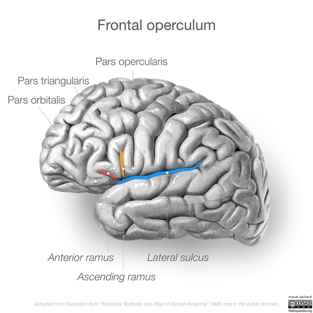
Pars orbitalis
Citation, DOI, disclosures and article data
At the time the article was created Callum Smith had no recorded disclosures.
View Callum Smith's current disclosuresAt the time the article was last revised Craig Hacking had no recorded disclosures.
View Craig Hacking's current disclosures- Brodmann's area 47/12
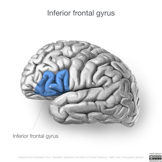
The pars orbitalis refers to the most rostral portion of the inferior frontal gyrus in the frontal lobe of the brain. It is one of three parts that make up the inferior frontal gyrus along with the pars triangularis and pars opercularis 4-6. It plays a role in the language processing network of the brain.
On this page:
Gross anatomy
Relations
The pars orbitalis lies ventral to the anterior horizontal ramus (a.k.a. anterior ramus) of the lateral sulcus (a.k.a. Sylvian fissure). It extends along the ventral most part of the lateral surface of the frontal lobe as far as the caudal segment of the lateral orbital sulcus 4-6.
Blood supply
It is supplied by the frontal branches of the middle cerebral artery.
Radiographic features
Radiographically the pars orbitalis can be identified as the most rostral portion of the inferior frontal gyrus which takes on a characteristic “M” configuration. This is also referred to as the M sign when identifying the inferior frontal gyrus 3.
Function
Although the pars orbitalis is not defined by cytoarchitecture, it is closely affiliated to Brodmann’s areas 47/12 1. Traditionally not considered to be a core language area, new studies suggest in the dominant hemisphere it plays a more significant role in language processing with its involvement in semantic processing, phonological processing and syntax 1. The function of the same cortical region in the non-dominant hemisphere is not as well documented but has been shown to be involved in behavioural and motor inhibition and deductive reasoning 2.
References
- 1. De Carli D, Garreffa G, Colonnese C, Giulietti G, Labruna L, Briselli E, Ken S, Macrì MA, Maraviglia B. Identification of activated regions during a language task. Magnetic resonance imaging. 25 (6): 933-8. doi:10.1016/j.mri.2007.03.031 - Pubmed
- 2. Wildgruber D, Riecker A, Hertrich I, Erb M, Grodd W, Ethofer T, Ackermann H. Identification of emotional intonation evaluated by fMRI. NeuroImage. 24 (4): 1233-41. doi:10.1016/j.neuroimage.2004.10.034 - Pubmed
- 3. M. Wagner, A. Jurcoane, E. Hattingen. The U Sign: Tenth Landmark to the Central Region on Brain Surface Reformatted MR Imaging. American Journal of Neuroradiology. 34 (2): 323. doi:10.3174/ajnr.A3205 - Pubmed
- 4. Michael Petrides. Neuroanatomy of Language Regions of the Human Brain.(2014) ISBN: 9780124059313
- 5. Juergen K. Mai, George Paxinos. The Human Nervous System.(2012) ISBN: 9780080921303
- 6. Stephan Ulmer, Olav Jansen. fMRI. ISBN: 9783642343421
Incoming Links
Related articles: Anatomy: Brain
-
brain
- grey matter
- white matter
-
cerebrum
-
cerebral hemisphere (telencephalon)
- cerebral lobes and gyri
- frontal lobe
- parietal lobe
-
occipital lobe
- occipital pole
- lingual gyrus
- fusiform gyrus (Brodmann area 37)
- calcarine (visual) cortex
- cuneus
- temporal lobe
- basal forebrain
- limbic system
- insula
-
cerebral sulci and fissures (A-Z)
- calcarine fissure
- callosal sulcus
- central (Rolandic) sulcus
- cingulate sulcus
- collateral sulcus
- inferior frontal sulcus
- inferior occipital sulcus
- inferior temporal sulcus
- interhemispheric fissure
- intraparietal sulcus
- lateral (Sylvian) sulcus
- lateral occipital sulcus
- marginal sulcus
- occipitotemporal sulcus
- olfactory sulcus
- paracentral sulcus
- paraolfactory sulcus
- parieto-occipital fissure
- posterior parolfactory sulcus
- precentral sulcus
- preoccipital notch
- postcentral sulcus
- rhinal sulcus
- rostral sulcus
- subparietal sulcus
- superior frontal sulcus
- superior occipital sulcus
- superior temporal sulcus
- cortical histology
- cerebral lobes and gyri
- white matter tracts
- deep grey matter
-
pituitary gland
- posterior pituitary and stalk (part of diencephalon)
- anterior pituitary
- inferior hypophyseal arterial circle
- diencephalon
-
cerebral hemisphere (telencephalon)
-
brainstem
- midbrain (mesencephalon)
- pons (part of metencephalon)
- medulla oblongata (myelencephalon)
- white matter
-
grey matter
- non-cranial nerve
-
cranial nerve nuclei
- oculomotor nucleus
- Edinger-Westphal nucleus
- trochlear nucleus
- motor nucleus of CN V
- mesencephalic nucleus of CN V
- main sensory nucleus of CN V
- spinal nucleus of CN V
- abducent nucleus
- facial nucleus
- superior salivatory nucleus
- cochlear nuclei
- vestibular nuclei
- inferior salivatory nucleus
- solitary tract nucleus
- ambiguus nucleus
- dorsal vagal motor nucleus
- hypoglossal nucleus
-
cerebellum (part of metencephalon)
- vermis
- cerebellar hemisphere
- cerebellar peduncles
- cranial meninges (meninx primitiva)
- CSF spaces
-
cranial nerves (mnemonic)
- olfactory nerve (CN I)
- optic nerve (CN II)
- oculomotor nerve (CN III)
- trochlear nerve (CN IV)
- trigeminal nerve (CN V) (mnemonic)
- abducens nerve (CN VI)
- facial nerve (CN VII) (segments mnemonic | branches mnemonic)
-
vestibulocochlear nerve (CN VIII)
- vestibular ganglion (Scarpa's ganglion)
- glossopharyngeal nerve (CN IX)
- vagus nerve (CN X)
- spinal accessory nerve (CN XI)
- hypoglossal nerve (CN XII)
- functional neuroanatomy
- CNS development
- cerebral vascular supply
- arteries
- vascular territories
-
circle of Willis
- internal carotid artery (ICA) (segments)
- vertebral artery
-
normal variants
- intracranial arterial fenestration
- internal carotid artery (ICA)
- anterior cerebral artery (ACA)
- middle cerebral artery (MCA)
- posterior cerebral artery (PCA)
- basilar artery
- persistent carotid-vertebrobasilar artery anastomoses (mnemonic)
- vertebral artery
- ophthalmic artery
-
cerebral venous system
-
dural venous sinuses
- basilar venous plexus
- cavernous sinus (mnemonic)
- clival diploic veins
- inferior petro-occipital vein
- inferior petrosal sinus
- inferior sagittal sinus
- intercavernous sinus
- internal carotid artery venous plexus of Rektorzik
- jugular bulb
- marginal sinus
- occipital sinus
- sigmoid sinus
- sphenoparietal sinus
- straight sinus
- superior petrosal sinus
- superior sagittal sinus
- torcula herophili
- transverse sinus
-
cerebral veins
-
superficial veins of the brain
- superior cerebral veins (superficial cerebral veins)
- inferior cerebral veins
- superficial middle cerebral vein
- superior anastomotic vein (of Trolard)
- inferior anastomotic vein (of Labbe)
-
superficial veins of the brain
-
deep veins of the brain
- great cerebral vein (of Galen)
- venous circle of Trolard
- normal variants
-
dural venous sinuses
- arteries
- glymphatic pathway


 Unable to process the form. Check for errors and try again.