
MRI protocol for the assessment of perianal fistulas is a group of MRI sequences put together to asses the extension and anatomic relationships of inflammatory fistulas to the anal sphincters, helping to plan surgical management and monitor treatment response.
NB: This article is intended to outline some general principles of protocol design. The specifics will vary depending on MRI hardware and software, radiologist's and referrer's preferences, institutional protocols, patient factors (e.g. allergy) and time constraints.
Sequences
A good protocol for this purpose would include:
-
fine 3 mm slices
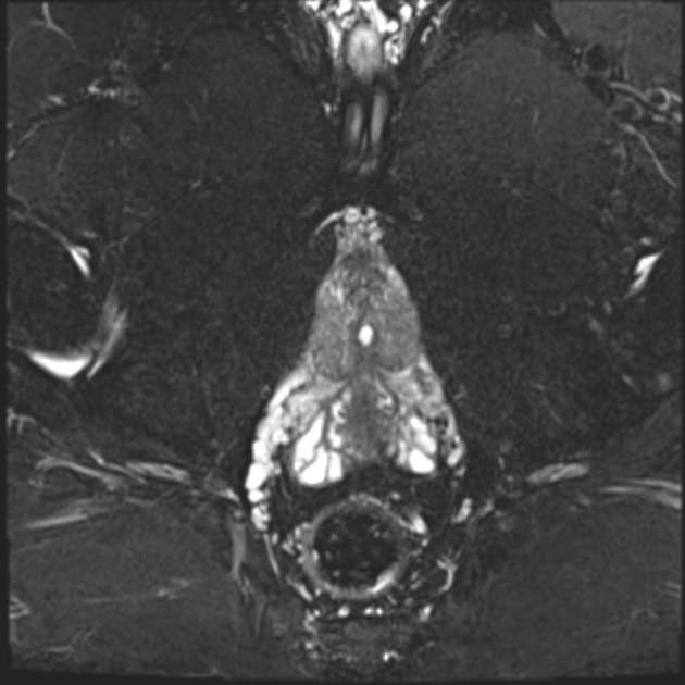
axial and coronal T2 FS or STIR
axial and coronal T2 are helpful in the delineation of sphincter anatomy
axial and coronal T1 FS with gadolinium-based intravenous contrast material
axial T1
Some authors recommend 1:
short-inversion-time inversion recovery (STIR)
gradient-echo T1-weighted with or without gadolinium-based intravenous contrast material
spin-echo T1-weighted
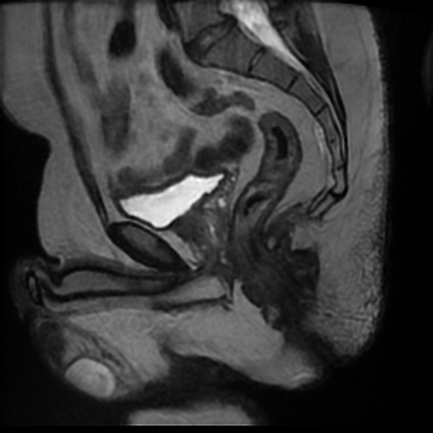
spin-echo T2-weighted with saline instillation (MR fistulography)
A novel method is the use of volume MRI sequences (more routinely used in neuro and musculoskeletal MRI). The benefits are reduced imaging times and isotropic data allowing multiplanar reformatting while maintaining comparable contrast resolution.


 Unable to process the form. Check for errors and try again.