
Petrosquamous fissure
Citation, DOI, disclosures and article data
At the time the article was created Nafisa Shakir Batta had no recorded disclosures.
View Nafisa Shakir Batta's current disclosuresAt the time the article was last revised Francis Deng had no recorded disclosures.
View Francis Deng's current disclosures- Fissura petrosquamosa
- Petrosquamous suture
- Petrosquamosal suture
- Petrosquamosal fissure
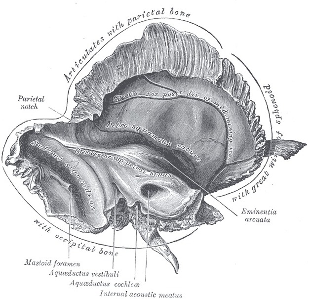
The petrosquamous fissure is the obliquely oriented fissure between the medial petrous part and lateral squamous part of the temporal bone.
Gross anatomy
The anterior (ventral) petrosquamous fissure is the medial continuation of the tympanosquamous fissure and petrotympanic fissure (together forming a Y configuration). It is oriented obliquely toward the greater wing of sphenoid where it splits into the sphenosquamosal suture and sphenopetrosal suture. This part of the fissure is posterior to the squamous part of temporal bone that is medial to the mandibular fossa, and anterior to the petrous part of temporal bone that is an inferior projection of the tegmen tympani known as the intertympanosquamosal crest or crista tegmentalis 2.
The middle petrosquamous fissure is difficult to delineate in the adult. On the intratympanic side, the fissure may be related to the cog 2. On the endocranial side, the fissure may show a groove for the petrosquamous sinus, a tributary of the sigmoid sinus 2.
At the posterior petrosquamous fissure, the mastoid tegmen projects inferiorly due to developmental apposition between squamous and petrosal parts, forming Koerner septum internally 2. Externally, the posterior petrosquamous fissure is also known as the squamomastoid suture.
ADVERTISEMENT: Supporters see fewer/no ads
Radiographic features
CT
The anterior part of the fissure is seen on axial views as an anteromedially oriented cleft extending from the mandibular fossa toward the greater wing of the sphenoid 1.
The middle part of the fissure may be seen on coronal views as a tiny defect in the tegmen tympani 1.
The posterior part of the fissure is seen as a vertically oriented site of apposition on the mastoid surface between the squamous part (squamomastoid) and petrosal part (petromastoid) 2.
The Koerner septum is a useful landmark for approximating the orientation of the petrosquamous fissure.
References
- 1. Som PM, Curtin HD. Head and Neck Imaging. (2011) ISBN: 9780323053556
- 2. Virapongse C, Kirchner JC, Sasaki C, Shapiro M. Computed tomography of Körner's septum and petrosquamosal suture. (1986) Archives of otolaryngology--head & neck surgery. 112 (1): 81-7. doi:10.1001/archotol.1986.03780010083016 - Pubmed
Incoming Links
Related articles: Anatomy: Head and neck
- skeleton of the head and neck
-
cranial vault
- scalp (mnemonic)
- fontanelle
-
sutures
- calvarial
- facial
- frontozygomatic suture
- frontomaxillary suture
- frontolacrimal suture
- frontonasal suture
- temporozygomatic suture
- zygomaticomaxillary suture
- parietotemporal suture (parietomastoid suture)
- occipitotemporal suture (occipitomastoid suture)
- sphenofrontal suture
- sphenozygomatic suture
- spheno-occipital suture (not a true suture)
- lacrimomaxillary suture
- nasomaxillary suture
- internasal suture
- basal/internal
- skull landmarks
- frontal bone
- temporal bone
- parietal bone
- occipital bone
- skull base (foramina)
-
facial bones
- midline single bones
- paired bilateral bones
- cervical spine
- hyoid bone
- laryngeal cartilages
-
cranial vault
- muscles of the head and neck
- muscles of the tongue (mnemonic)
- muscles of mastication
-
facial muscles
- epicranius muscle
- circumorbital and palpebral muscles
- nasal muscles
-
buccolabial muscles
- elevators, retractors and evertors of the upper lip
- levator labii superioris alaeque nasalis muscle
- levator labii superioris muscle
- zygomaticus major muscle
- zygomaticus minor muscle
- levator anguli oris muscle
- malaris muscle
- risorius muscle
- depressors, retractors and evertors of the lower lip
- depressor labii inferioris muscle
- depressor anguli oris muscle
- mentalis muscle
- compound sphincter
-
orbicularis oris muscle
- incisivus labii superioris muscle
- incisivus labii inferioris muscle
-
orbicularis oris muscle
- muscle of mastication
- modiolus
- elevators, retractors and evertors of the upper lip
- muscles of the middle ear
- orbital muscles
- muscles of the soft palate
- pharyngeal muscles
- suprahyoid muscles
- infrahyoid muscles
- intrinsic muscles of the larynx
- muscles of the neck
- platysma muscle
- longus colli muscle
- longus capitis muscle
- scalenus anterior muscle
- scalenus medius muscle
- scalenus posterior muscle
- scalenus pleuralis muscle
- sternocleidomastoid muscle
-
suboccipital muscles
- rectus capitis posterior major muscle
- rectus capitis posterior minor muscle
- obliquus capitis superior muscle
- obliquus capitis inferior muscle
- accessory muscles of the neck
- deep cervical fascia
-
deep spaces of the neck
- anterior cervical space
- buccal space
- carotid space
- danger space
- deep cervical fascia
- infratemporal fossa
- masticator space
- parapharyngeal space
- stylomandibular tunnel
- parotid space
- pharyngeal (superficial) mucosal space
- perivertebral space
- posterior cervical space
- pterygopalatine fossa
- retropharyngeal space
- suprasternal space (of Burns)
- visceral space
- surgical triangles of the neck
- orbit
- ear
- paranasal sinuses
- upper respiratory tract
- viscera of the neck
- blood supply of the head and neck
-
arterial supply
-
common carotid artery
- carotid body
- carotid bifurcation
- subclavian artery
- variants
-
common carotid artery
- venous drainage
-
arterial supply
- innervation of the head and neck
-
cranial nerves
- olfactory nerve (CN I)
- optic nerve (CN II)
- oculomotor nerve (CN III)
- trochlear nerve (CN IV)
-
trigeminal nerve (CN V) (mnemonic)
- trigeminal ganglion
- ophthalmic division
- maxillary division
- mandibular division
- abducens nerve (CN VI)
- facial nerve (CN VII)
-
vestibulocochlear nerve (CN VIII)
- vestibular ganglion (Scarpa's ganglion)
- glossopharyngeal nerve (CN IX)
- vagus nerve (CN X)
- (spinal) accessory nerve (CN XI)
- hypoglossal nerve (CN XII)
- parasympathetic ganglia of the head and neck
- cervical sympathetic ganglia
- greater occipital nerve
- third occipital nerve
-
cervical plexus
- muscular branches
- longus capitis
- longus colli
- scalenes
- geniohyoid
- thyrohyoid
-
ansa cervicalis
- omohyoid (superior and inferior bellies separately)
- sternothyroid
- sternohyoid
- phrenic nerve
- contribution to the accessory nerve (CN XI)
- cutaneous branches
- muscular branches
- brachial plexus
- pharyngeal plexus
-
cranial nerves
- lymphatic drainage of the head and neck
- embryological development of the head and neck


 Unable to process the form. Check for errors and try again.