
Pituitary stalk abnormal enhancement (differential)
Citation, DOI, disclosures and article data
Citation:
Gaillard F, Glick Y, Bodlak C, et al. Pituitary stalk abnormal enhancement (differential). Reference article, Radiopaedia.org (Accessed on 13 Mar 2025) https://doi.org/10.53347/rID-1891
rID:
1891
Article created:
Disclosures:
At the time the article was created Frank Gaillard had no recorded disclosures.
View Frank Gaillard's current disclosures
Last revised:
Disclosures:
At the time the article was last revised Yaïr Glick had no recorded disclosures.
View Yaïr Glick's current disclosures
Revisions:
13 times, by
11 contributors -
see full revision history and disclosures
Systems:
Synonyms:
- Abnormal enhancement / bulkiness of the pituitary infundibulum
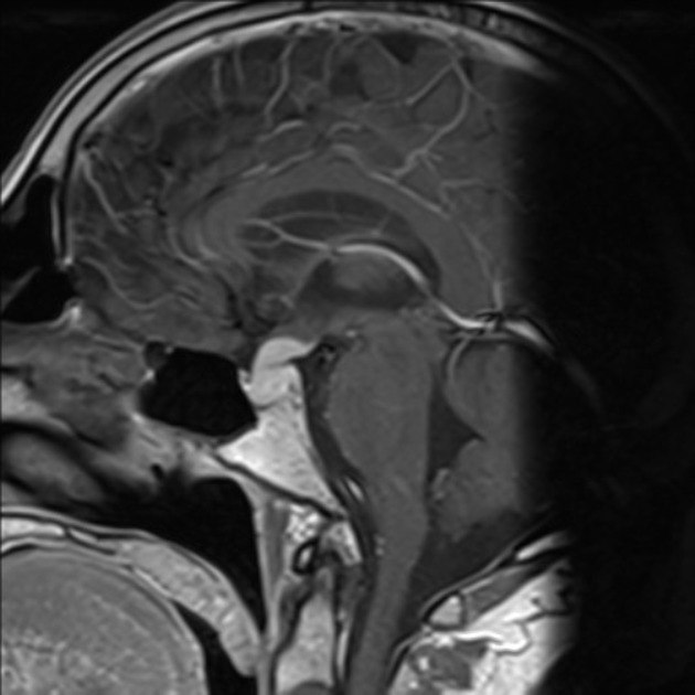
Abnormal nodular enhancement of the pituitary stalk can be seen in a number of entities.
Differential diagnosis
-
tumors
- germinoma
- craniopharyngioma
- hypothalamic glioma
- pituitary lymphoma
- pituicytoma
- granular cell tumor of the pituitary (pituitary choristoma)
- pilocytic astrocytoma of the neurohypophysis (infundibuloma)
- pituitary metastases
- CNS hemangioblastoma related to Von Hippel-Lindau disease
-
infection
- CNS tuberculosis
- sequelae of meningitis
- cellular infiltrates
References
- 1. Loevner LA. Brain imaging, case review. Mosby Inc. (2008) ISBN:032303179X. Read it at Google Books - Find it at Amazon
- 2. Batista RL, Ramos LS, Cescato VA, Musolino NR, Borba CG, Silva GO, Moreno LH, Cunha Neto MB. Thickened Pituitary Stalk Associated with a Mass in the Sphenoidal Sinus: An Alarm to Suspect Hypophysitis by Immunoglobulin G4?. (2015) International archives of otorhinolaryngology. 19 (3): 273-6. doi:10.1055/s-0034-1397333 - Pubmed
- 3. Lonser R, Butman J, Kiringoda R, Song D, Oldfield E. Pituitary Stalk Hemangioblastomas in Von Hippel-Lindau Disease. J Neurosurg. 2009;110(2):350-3. doi:10.3171/2008.4.17532 - Pubmed
Incoming Links
Articles:
Related articles: Pituitary region masses
-
general reading
- pituitary gland anatomy
- pituitary MRI - an approach
-
pituitary region masses
- most common pituitary region masses
- solid and enhancing pituitary region mass
- mixed cystic and solid pituitary region mass
- mostly/purely cystic pituitary region masses
- purely intrasellar pituitary mass
- pituitary region mass with intrinsic high T1 signal
- abnormal enhancement/bulkiness of the pituitary infundibulum
- enlarged sella turcica
- mnemonic: SATCHMO
- history of imaging the pituitary region
-
pathology
-
pituitary tumors
- pituitary adenoma (commonest in the adult population)
- pituitary carcinoma
- pituitary lymphoma
- meningioma
- craniopharyngioma
- optic pathway glioma
- germinoma
- chordoma
- dermoid (CNS) / epidermoid / intracranial teratoma
- pituicytoma
- spindle cell oncocytomas
- pituitary metastases
- granular cell tumor of the pituitary (pituitary choristoma)
- pilocytic astrocytoma of the neurohypophysis (infundibuloma)
- cellular infiltrates
- other lesions
- anterior circulation berry aneurysm
- hamartoma (tuber cinereum hamartoma)
- Rathke cleft cyst
- intracranial lipoma
- sphenoid sinus mucocoele
- pituitary abscess
- pituitary stone
-
pituitary tumors


 Unable to process the form. Check for errors and try again.