
Placental evaluation with MRI is a problem-solving technique that can be used if ultrasound evaluation is insufficient or confusing. Even if the placenta is not the main point of evaluation, it is useful to understand the appearance of the placenta on obstetric imaging for other causes.
On this page:
Technique
Placental evaluation with MRI is not generally performed in the first trimester due to a theoretical risk of MRI to the embryo.
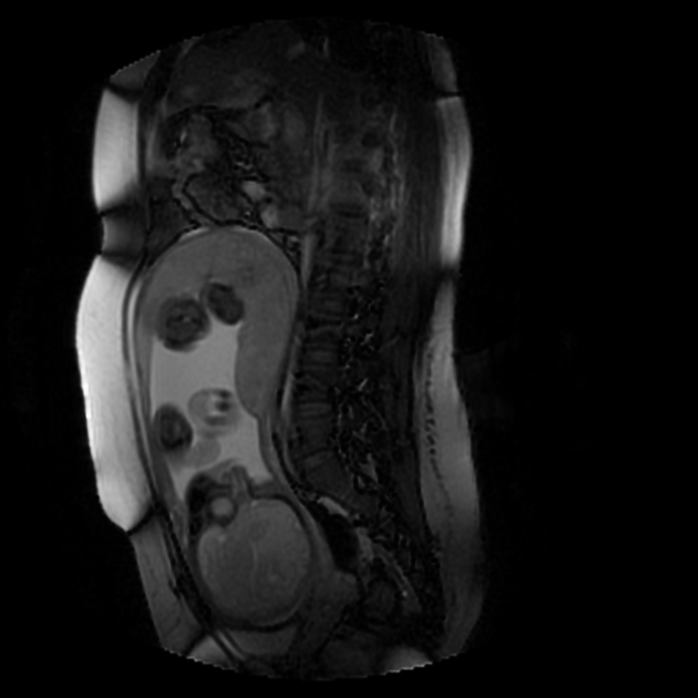
Placental evaluation relies on two main sequences (acquired in all three planes):
- single-shot fast spin-echo (SSFSE)
- or half-acquisition turbo spin-echo (HASTE)
- balanced steady-state free procession (bSSFP)
Additional planes may be useful for evaluation, as needed.
An axial T1 fat-suppressed may be added to evaluate for blood products. Diffusion-weighted sequences have also been used by some to evaluate the placental-myometrial interface.
Intravenous gadolinium contrast is not generally used out of potential risk to the fetus.
Radiographic features
Normal appearance
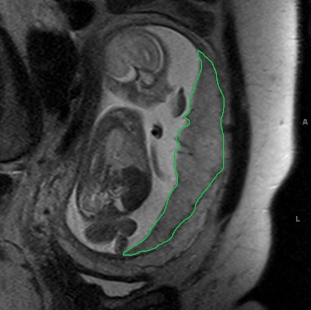
The placenta is a 2-4 cm thick pancake-shaped structure attached to either the anterior or posterior uterine cavity.
The placenta's imaging appearance changes during gestation:
- 19-23 weeks
- T2: homogeneous signal intensity
- 24-31 weeks
- placenta becomes lobulated
- septae appear between the placental lobules
- T2: increasingly heterogeneous signal intensity
Evaluation of abnormal appearance
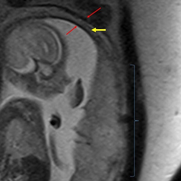
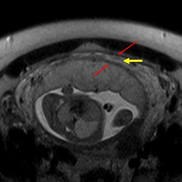
An abnormal appearance of the placental implantation relies on disruption of the normal T2 trilayered appearance of gravid myometrium (central hyperintense vascular layer between two hypointense layers) 2. Abnormal placentation is not reliably assessed prior to 24 weeks gestational age 4.
Differential diagnosis
Placental MRI can be very useful in confirming and characterizing of disorders of abnormal placental villous adherence:
- placenta accreta
- placenta increta
- placenta percreta (the bladder should be mildly distended if evaluating for percreta on MRI)
Other situations in which it may be useful
- abnormal placental location
- delineating bilobed placenta or accessory placenta
- placental vascular anomalies
Practical points
- mild placental lobulation and myometrial thinning can be seen in normal placental implantation
- placental heterogeneity normally increases with gestational age
- abnormal placental villous adherence should be confirmed in two planes


 Unable to process the form. Check for errors and try again.