
Popliteal venous aneurysms are rare than those of the popliteal artery and are mostly asymptomatic. However, due to the disturbance of the venous blood flow, they can lead to potentially life-threatening consequences, such as deep vein thrombosis (DVT) and pulmonary embolism (PE).
On this page:
Epidemiology
Popliteal venous aneurysms are uncommon, during duplex ultrasound scans an incidence of 0.1-0.2% was observed, with a slight left-sided and female predominance 1.
Clinical presentation
mostly asymptomatic, discovered when sequelae such as deep vein thrombosis or pulmonary embolism develop
resistance in the popliteal fossa
Pathology
The pathogenesis is poorly understood. Congential weakness of the venous wall, inflammation, trauma, and hemodynamic changes have all been suggested as causative factors 2.
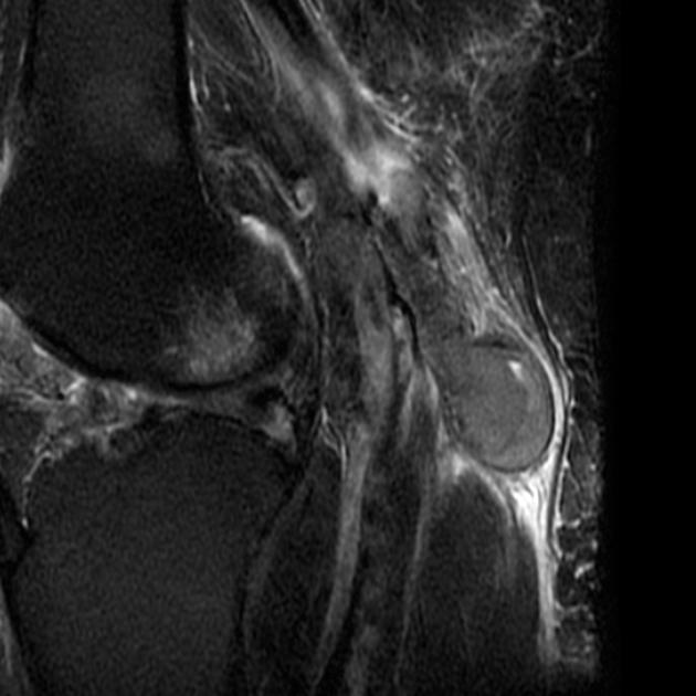
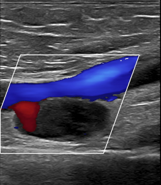
Radiographic features
Phlebography
Used to be the gold standard, no longer routinely used 1-3.
Ultrasound
The initial imaging modality of choice, sensitive, with color Doppler also allows dynamic evaluation of blood flow.
CT/MRI
CT and MRI angiography are also capable of demonstrating the pathology 4.
Treatment and prognosis
Open surgical repair is the mainstay of the treatment 1,4.
Complications
deep vein thrombosis
pulmonary embolism
Differential diagnosis
popliteal cyst (Baker's cyst)
miscellaneous cystic masses in the same region such as synovial sarcoma


 Unable to process the form. Check for errors and try again.