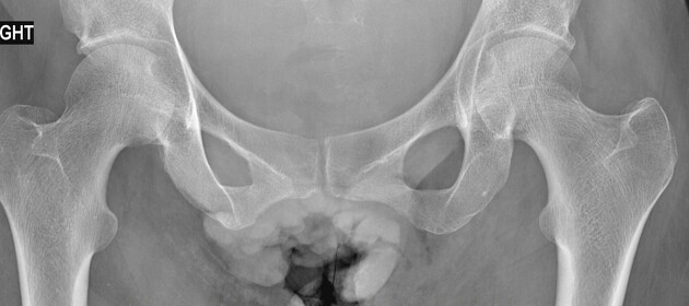
Primary vulval cancer is a rare gynecological malignancy that originates from the vulva.
On this page:
Epidemiology
It accounts for ~3-5% of female genital tract malignancies and typically presents in postmenopausal patients peaking around the age of 65-70 years of age 1.
Pathology
The commonest histological type by far is squamous cell carcinoma which account of ~80-85% of cases 1. The tumor commonly involves the labia majora and minora.
Risk factors
- vulval intraepithelial neoplasia: considered a precancerous condition
Radiographic features
MRI
Particularly useful in accurately assessing the size (up to ~80 accurate 3) of vulval lesion and assessing groin lymph node metastasis
Signal characterisitcs include:
- T1: low 4 to intermediate 1 signal
- T2: typically intermediate 4 to high 1 signal
Staging
The FIGO staging system is commonly adopted: see vulval cancer staging
Differential diagnosis
Considerations (particularly when lesions are large) include:
- vaginal carcinoma with vulval extension
- cervical carcinoma with vulval extension


 Unable to process the form. Check for errors and try again.