
Renal vein
Citation, DOI, disclosures and article data
At the time the article was created Henry Knipe had no recorded disclosures.
View Henry Knipe's current disclosuresAt the time the article was last revised Craig Hacking had the following disclosures:
- Philips Australia, Paid speaker at Philips Spectral CT events (ongoing)
These were assessed during peer review and were determined to not be relevant to the changes that were made.
View Craig Hacking's current disclosures- Renal veins
- Renal vein anatomy
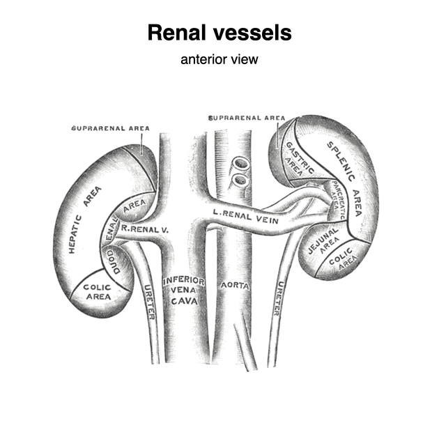
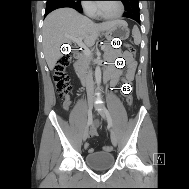
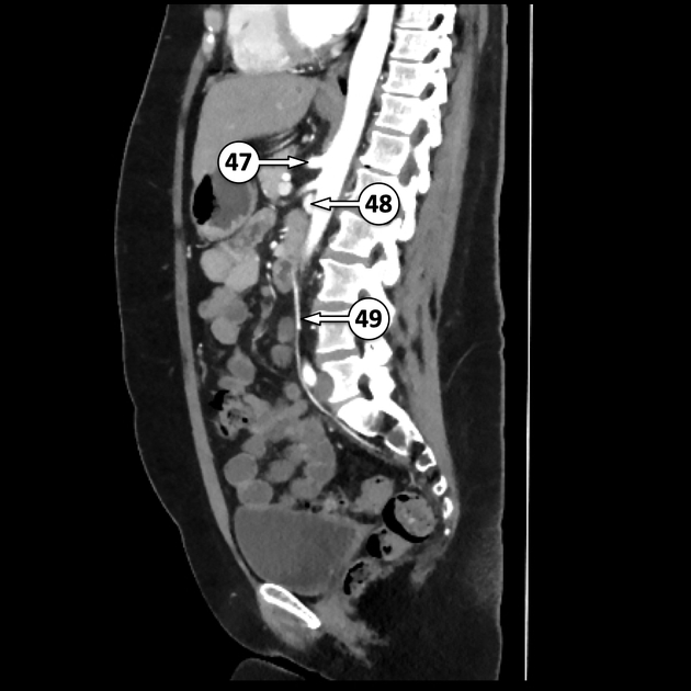
The renal veins are asymmetric paired retroperitoneal veins that drain the kidneys.
On this page:
Gross anatomy
Course
The renal vein is formed by the union of two-to-three renal parenchymal veins in the renal sinus. It emerges from the renal hilum anterior to the renal artery and drains into the inferior vena cava at the level of L2.
The left renal vein is much longer, at 6-7 cm, than the right renal vein, at 3-4 cm, but they have a similar calibre (~1.2 cm). The left renal vein courses anteriorly to the abdominal aorta.
Tributaries
Left renal vein
- left gonadal vein
- left inferior phrenic vein
- left adrenal vein
- small branches from kidney capsule, proximal ureter and renal pelvis
Right renal vein
- small branches from kidney capsule, proximal ureter and renal pelvis
Variant anatomy
See article: renal vein anomalies.
Related pathology
References
- 1. Geoffrey D. Rubin, Neil M. Rofsky. CT and MR Angiography. (2009) ISBN: 9780781745253 - Google Books
- 2. Michael Schünke, Erik Schulte, Udo Schumacher. Thieme Atlas of Anatomy. (2006) ISBN: 9783131420916 - Google Books
- 3. Mauro MA, Murphy KP, Thomson KR et-al. Image-Guided Interventions: Expert Radiology Series. Saunders. ISBN:B00E6GB08K. Read it at Google Books - Find it at Amazon
Incoming Links
- Adrenal gland
- Surgical splenorenal shunt
- Superior mesenteric artery
- Vascular compression disorders
- Splenic vein
- Common iliac vein
- Para-aortic lymph nodes
- Adrenal veins
- Superior mesenteric artery compression disorders
- Testes
- Pelvic congestion syndrome
- Tributaries of the inferior vena cava (mnemonic)
- Kidneys
- Threads and streaks sign
- Abernethy malformation
- Spontaneous splenorenal shunt
- Renal artery
- RENAL nephrometry scoring system
- Ovary
- Ascending lumbar communicant vein
- Renal vessels (Gray's illustrations)
- Aberrant extra renal artery and vein arising from common iliac vessels
- Renal artery aneurysm causing focal hydronephrosis
- Transposition of inferior vena cava
- Pelviureteric junction obstruction by accessory renal vein
- Renal cell carcinoma
- Cardiac arrest (CT)
- Circumaortic left renal vein
- Metastatic locally invasive renal cell carcinoma
- Truncal venous development (Gray's illustration)
- Retroaortic left renal vein
- Hepatic hydatid cyst and Nutcracker phenomenon
- Adrenal cortical carcinoma
- Left renal vein thrombosis
- Nutcracker phenomenon
- Left testicular varicocele - nutcracker phenomenon
Related articles: Anatomy: Abdominopelvic
- skeleton of the abdomen and pelvis
- muscles of the abdomen and pelvis
- spaces of the abdomen and pelvis
- anterior abdominal wall
- posterior abdominal wall
- abdominal cavity
- pelvic cavity
- perineum
- abdominal and pelvic viscera
- gastrointestinal tract
- spleen
- hepatobiliary system
-
endocrine system
-
adrenal gland
- adrenal vessels
- chromaffin cells
- variants
- pancreas
- organs of Zuckerkandl
-
adrenal gland
-
urinary system
-
kidney
- renal pelvis
- renal sinus
- avascular plane of Brodel
-
variants
- number
- fusion
- location
- shape
- ureter
- urinary bladder
- urethra
- embryology
-
kidney
- male reproductive system
-
female reproductive system
- vulva
- vagina
- uterus
- adnexa
- Fallopian tubes
- ovaries
- broad ligament (mnemonic)
- variant anatomy
- embryology
- blood supply of the abdomen and pelvis
- arteries
-
abdominal aorta
- inferior phrenic artery
- coeliac artery
- superior mesenteric artery
- middle suprarenal artery
- renal artery (variant anatomy)
- gonadal artery (ovarian artery | testicular artery)
- inferior mesenteric artery
- lumbar arteries
- median sacral artery
-
common iliac artery
- external iliac artery
-
internal iliac artery (mnemonic)
- anterior division
- umbilical artery
- superior vesical artery
- obturator artery
- vaginal artery
- inferior vesical artery
- uterine artery
- middle rectal artery
-
internal pudendal artery
- inferior rectal artery
-
perineal artery
- posterior scrotal artery
- transverse perineal artery
- artery to the bulb
- deep artery of the penis/clitoris
- dorsal artery of the penis/clitoris
- inferior gluteal artery
- posterior division (mnemonic)
- variant anatomy
- anterior division
-
abdominal aorta
- portal venous system
- veins
- anastomoses
- arterioarterial anastomoses
- portal-systemic venous collateral pathways
- watershed areas
- arteries
- lymphatics
- innervation of the abdomen and pelvis
- thoracic splanchnic nerves
- lumbar plexus
-
sacral plexus
- lumbosacral trunk
- sciatic nerve
- superior gluteal nerve
- inferior gluteal nerve
- nerve to piriformis
- perforating cutaneous nerve
- posterior femoral cutaneous nerve
- parasympathetic pelvic splanchnic nerves
- pudendal nerve
- nerve to quadratus femoris and inferior gemellus muscles
- nerve to internal obturator and superior gemellus muscles
- autonomic ganglia and plexuses


 Unable to process the form. Check for errors and try again.