
Right middle lobe syndrome refers to chronic right middle lobe collapse, usually without an obstructing lesion (but not always). It is usually with associated bronchiectasis.
On this page:
Epidemiology
Right middle lobe syndrome is usually encountered in older adults, with a predilection for women (see Lady Windermere syndrome). It is also seen in children 1.
Clinical presentation
In most cases, patients are asymptomatic. Otherwise, a chronic cough can be a common symptom. Haemoptysis, chest pain, and dyspnoea have also been reported 1.
Pathology
Right middle lobe syndrome can be categorised into two types:
The underlying aetiology of right middle lobe syndrome remains poorly understood, but poor collateral ventilation, a relatively narrow ostium, and infection/inflammation are all thought to play a role 3. The histological processes identified in such cases include 1:
lymphoid hyperplasia
granulomatous inflammation
Mycobacterium avium complex infection (classic but found only in a minority of cases)
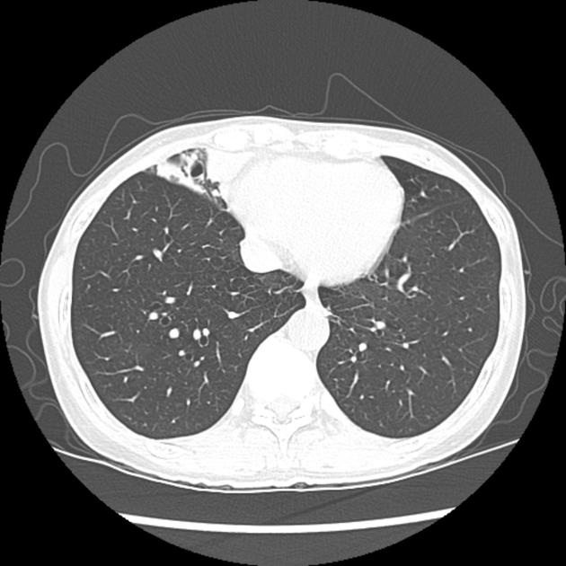
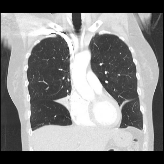
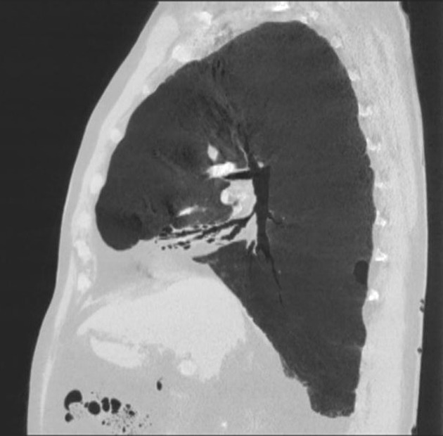
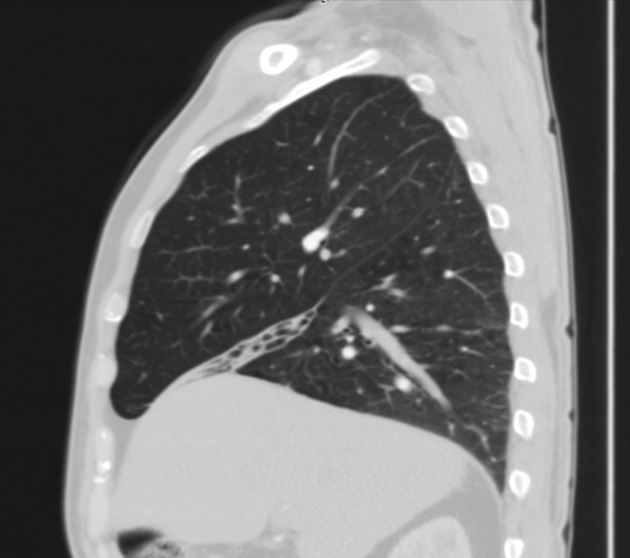
Radiographic features
The radiographic features of right middle lobe syndrome are a combination of:
History and etymology
Right middle lobe syndrome was first identified clinically in 1948 by Graham et al.


 Unable to process the form. Check for errors and try again.