
Schistosomiasis hepatic manifestations are a chronic result of the deposition of eggs into small portal venules, leading to periportal fibrosis and liver cirrhosis.
For a general overview, please refer to the main article on schistosomiasis.
On this page:
Epidemiology
Associations
Associated with an increased risk of hepatocellular carcinoma, particularly secondary to the S.Mansoni subtype
Clinical presentation
Usually, these patients will present with portal hypertension signs and symptoms, including splenomegaly, gastro-oesophageal varices, haematemesis, and ascites. Hepatic function is commonly preserved until late-stage disease 2.
Pathology
Living in the bowel lumen, schistosomes lay eggs in the mesenteric veins; thus, these eggs can reach the portal vein. Species S. mansoni and S. japonicum are the most common. An inflammatory reaction and consequent granulomatous response within the portal venules will lead to periportal fibrosis as a healing process 1-3.
Due to the difference in the size of the eggs, S. mansoni infection has its eggs deposited along the large portal veins of the hepatic hilum, whereas the S. japonicum infection has the eggs laying within the small peripheral portal veins 2.
Radiographic features
Imaging manifestations only occur late in the course of the chronic infection and there are some specific features related to the different distribution of the eggs depending on S. mansoni or S. japonicum infection.
Ultrasound
-
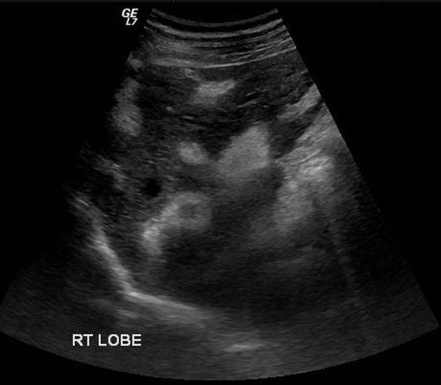
common features
irregular hepatic surface
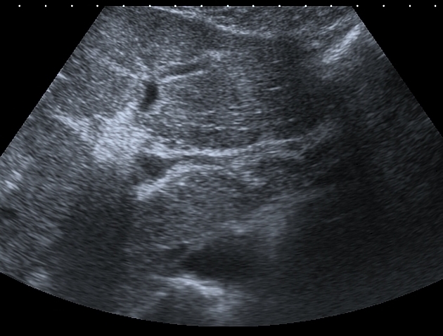
mosaic pattern: echogenic septa outlining polygonal areas of relatively normal liver parenchyma
septal fibrosis
-
splenomegaly
-
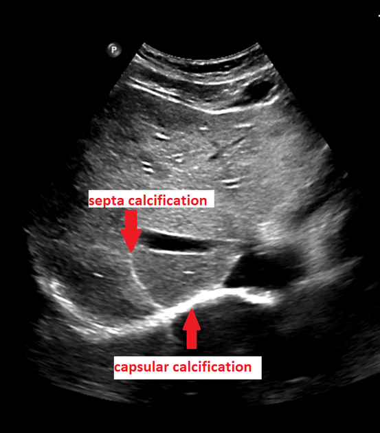
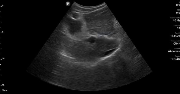
S. japonicum
septa and capsular calcifications
-
S. mansoni
-
portal veins wall thickening and increased echogenicity
“bull’s-eye” appearance: an anechoic portal vein surrounded by echogenic fibrous tissue
-
CT
-
common features
irregular hepatic contour
-
splenomegaly
-
S. japonicum
turtle back sign: calcified septa and fibrosis resembling a turtle carapace, considered pathognomonic
capsular calcification
periportal fat extending deep into the liver due to the parenchymal retraction
-
S. mansoni
low-attenuation surrounding the portal vein branches associated with marked contrast enhancement (periportal fibrosis) 1-2
eggs calcification is not seen as commonly as calcification of S.
japonicum eggs and consequently calcification along the portal tracts is not a common feature 2


 Unable to process the form. Check for errors and try again.