
Septal branches of the left anterior descending artery
Citation, DOI, disclosures and article data
Citation:
Edney G, Hacking C, Sharma R, Septal branches of the left anterior descending artery. Reference article, Radiopaedia.org (Accessed on 25 Mar 2025) https://doi.org/10.53347/rID-57664
rID:
57664
Article created:
5 Jan 2018,
Gabrielle Edney
Disclosures:
At the time the article was created Gabrielle Edney had no recorded disclosures.
View Gabrielle Edney's current disclosures
Last revised:
Disclosures:
At the time the article was last revised Craig Hacking had no recorded disclosures.
View Craig Hacking's current disclosures
Revisions:
5 times, by
3 contributors -
see full revision history and disclosures
Systems:
Sections:
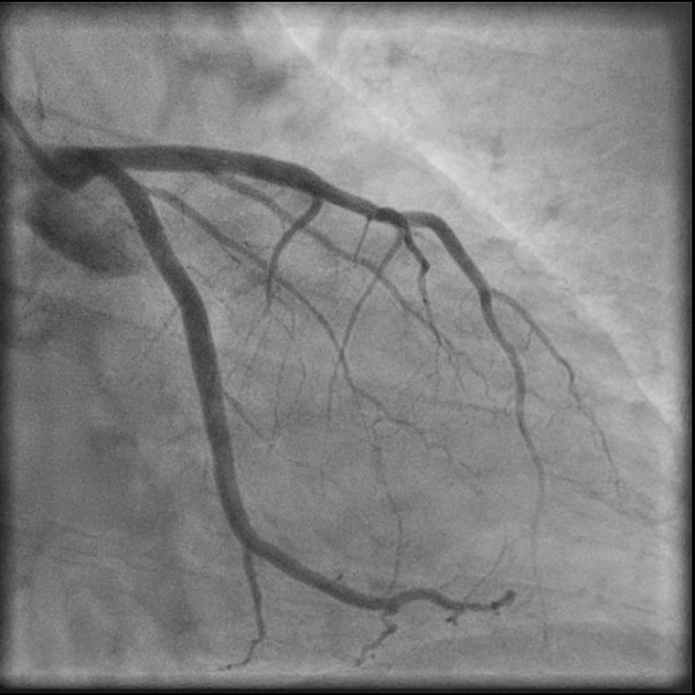
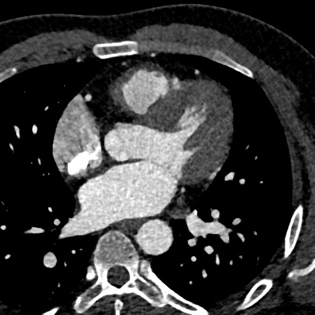
The septal branches of the left anterior descending artery supply blood flow to the interventricular septum of the heart.
Origin
These are right-sided branches (on axial CTCA) from the left anterior descending artery.
Supply
They provide the main blood supply to the anterior interventricular septum. A smaller posterior section is also supplied from the posterior descending artery instead (see coronary arterial dominance).
References
- 1. Wasilewski J, Roleder M et al. The role of septal perforators and "myocardial bridging effect" in atherosclerotic plaque distribution in the coronary artery disease. (2015) Polish journal of radiology. 80: 195-201. doi:10.12659/PJR.893227 - Pubmed
- 2. Mcminn. Last's Anatomy. ISBN: 9780729537520
Incoming Links
Related articles: Anatomy: Thoracic
- thoracic skeleton
- thoracic cage
- thoracic spine
- articulations
- muscles of the thorax
- diaphragm
- intercostal space
- intercostal muscles
- variant anatomy
- spaces of the thorax
- thoracic viscera
- lower respiratory tract
-
heart
- cardiac chambers
- heart valves
- cardiac fibrous skeleton
- innervation of the heart
- development of the heart
- cardiac wall
-
pericardium
- epicardium
- epicardial fat pad
- pericardial space
- oblique pericardial sinus
- transverse pericardial sinus
-
pericardial recesses
- aortic recesses
- pulmonic recesses
- postcaval recess
- pulmonary venous recesses
- pericardial ligaments
- myocardium
- endocardium
-
pericardium
- oesophagus
- thymus
- breast
- arterial supply of the thorax
-
thoracic aorta (development)
-
ascending aorta
-
aortic root
- aortic annulus
-
coronary arteries
- coronary arterial dominance
- myocardial segments
-
left main coronary artery (LMCA)
- ramus intermedius artery (RI)
-
circumflex artery (LCx)
- obtuse marginal branches (OM1, OM2, etc))
- Kugel's artery
-
left anterior descending artery (LAD)
- diagonal branches (D1, D2, etc)
- septal perforators (S1, S2, etc)
-
right coronary artery (RCA)
- conus artery
- sinoatrial nodal artery
- acute marginal branches (AM1, AM2, etc)
- inferior interventricular artery (PDA)
- posterior left ventricular artery (PLV)
- congenital anomalies
- sinotubular junction
-
aortic root
- aortic arch
- aortic isthmus
- descending aorta
-
ascending aorta
- pulmonary trunk
-
thoracic aorta (development)
- venous drainage of the thorax
- superior vena cava (SVC)
- inferior vena cava (IVC)
-
coronary veins
-
cardiac veins which drain into the coronary sinus
- great cardiac vein
- middle cardiac vein
- small cardiac vein
- posterior vein of the left ventricle
- vein of Marshall (oblique vein of the left atrium)
- anterior cardiac veins
- venae cordis minimae (smallest cardiac veins or thebesian veins)
-
cardiac veins which drain into the coronary sinus
- pulmonary veins
- bronchial veins
- thoracoepigastric vein
- lymphatics of the thorax
- innervation of the thorax


 Unable to process the form. Check for errors and try again.