
Superior sagittal sinus thrombosis (SSST) is the most common type of dural venous sinus thrombosis and is potentially devastating.
This article focuses on the specific features related to superior sagittal sinus thrombosis; please refer to the dural venous sinus thrombosis article for a general discussion.
Risk factors include pregnancy, dehydration, hypercoagulable states, and pancreatitis.
On this page:
Clinical presentation
As with all cerebral venous thrombosis, the presentation varies, ranging from completely asymptomatic to a rapid fulminant course with cerebral haemorrhage and death. Presentation includes:
headache: 53%, most common 1
seizures: 48%
hemi-, quadri-, or paraplegia: 48%
visual disturbances: 25%
nuchal rigidity: 18%
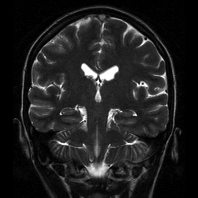
Radiographic features
Features are the same as those for other sinuses (please refer to dural venous sinus thrombosis).
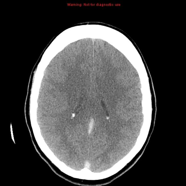
CT
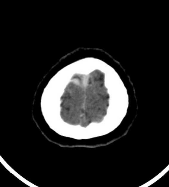
Unenhanced CT is usually the first imaging investigation performed, given the nonspecific clinical presentation in this case. When not associated with venous haemorrhage or infarction, it can be a subtle finding on CT images, relying on the hyperdensity of the sinus being identified. Potential findings include:
-
a potential pitfall is interpreting the distal superior sagittal sinus as being hyperdense near the torcular herophili
the walls at this location can be thick, measuring up to 2-3 mm
cerebral oedema: secondary to venous hypertension
cortical/cerebral swelling
unilateral or bilateral venous haemorrhage, including the cashew nut sign
With contrast administration, especially with a CT venogram, a filling defect in the sinus is sought. Multiplanar reformatted CT venography has been reported with a sensitivity of 95% for this diagnosis 4. In the superior sagittal sinus it is referred to as the empty delta sign. Signs of contrast CT include:
empty delta sign (is specific to the superior sagittal sinus)
prominent intramedullary veins
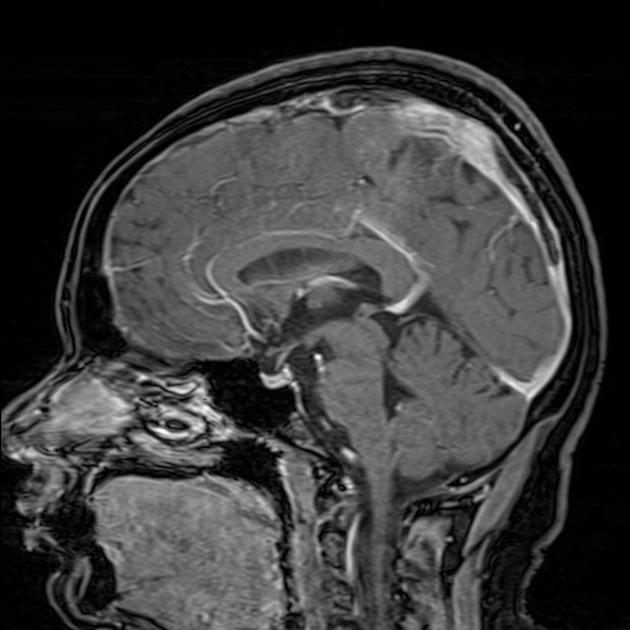
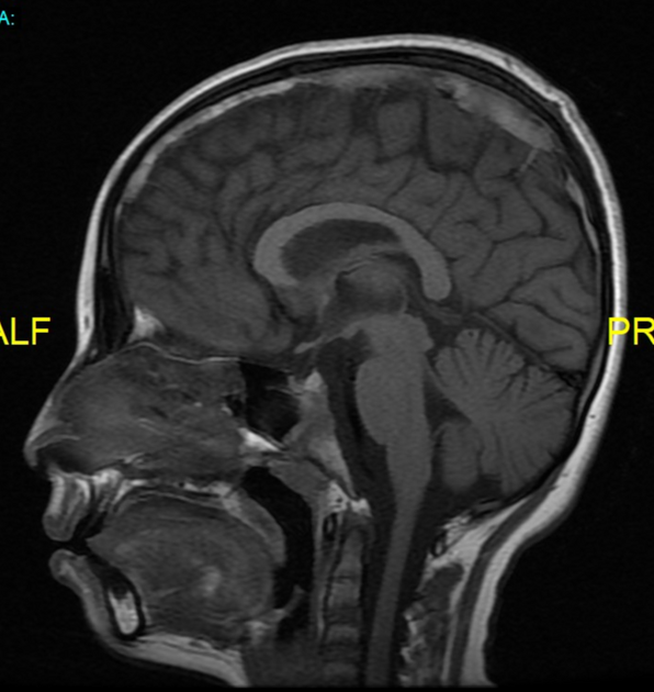
MRI
The clot acutely is isointense on T1 and hypointense on T2 (this can mimic a flow void), with subacute clot becoming hyperintense on T1. All the findings listed in the CT section can also be seen on MRI. MRV will demonstrate a lack of flow.
Treatment and prognosis
Management is the same as for other dural venous sinus thrombosis, please refer to the dural venous sinus thrombosis article for a general discussion.
Differential diagnosis
For high attenuating cerebral veins on a non-contrast CT scan, consider high haematocrit and/or haemoglobin levels 5.


 Unable to process the form. Check for errors and try again.