
Syntelencephaly, also known as middle interhemispheric variant (MIHV), is a mild subtype of holoprosencephaly that is characterised by an abnormal midline connection of the cerebral hemispheres between the posterior frontal and parietal regions.
On this page:
Epidemiology
Syntelencephaly is a congenital malformation, with no known racial or gender predilection.
Associations
Recognised associations include 1,2,4,5:
azygos anterior cerebral artery: usually present
dorsal cyst: seen, but much less frequently than in holoprosencephaly
-
cerebellar abnormalities
Clinical presentation
Patients with syntelencephaly present with a variety of deficits, particularly related to the involvement of the motor cortex. These include 2:
spasticity or hypotonia or dystonia
oromotor deficits affecting speech and feeding
Pathology
Unlike holoprosencephaly, to which syntelencephaly is believed to be related, the abnormality is usually confined to the posterior frontal and parietal lobes, often with the Sylvian fissure passing almost coronally over the vertex of the connected brain to join with the fissure from the other side.
Genetics
Mutations of the ZIC2 gene, on chromosome 13q32, thought to be important in regulating neural tube closure, have been implicated 1,2,5.
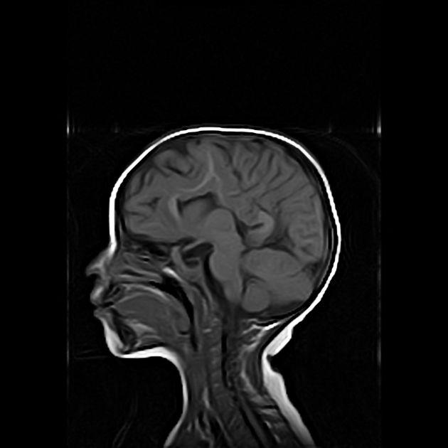
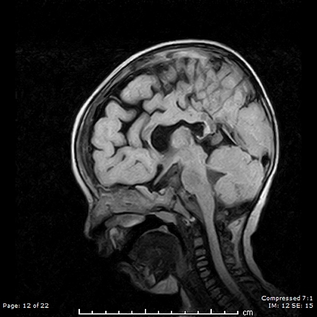
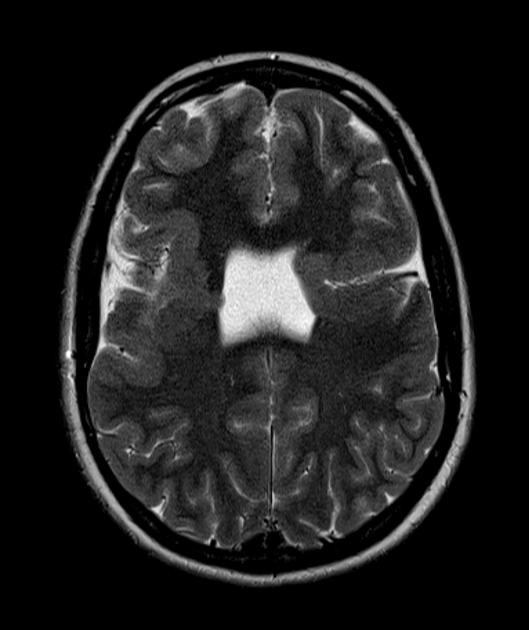
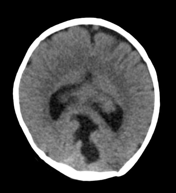
Radiographic features
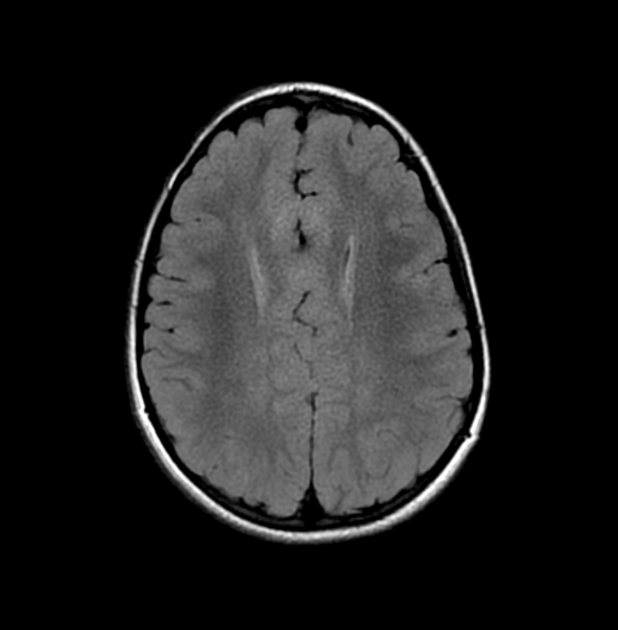
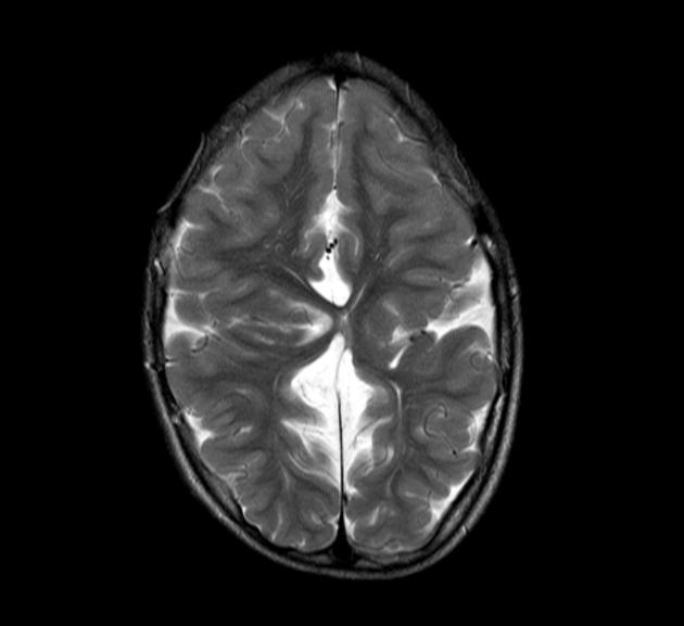
As with most cerebral structural congenital abnormalities, syntelencephaly is visible on all modalities, but in general is identified on antenatal ultrasound, and best characterised by MRI. Features that could be present are 1-5:
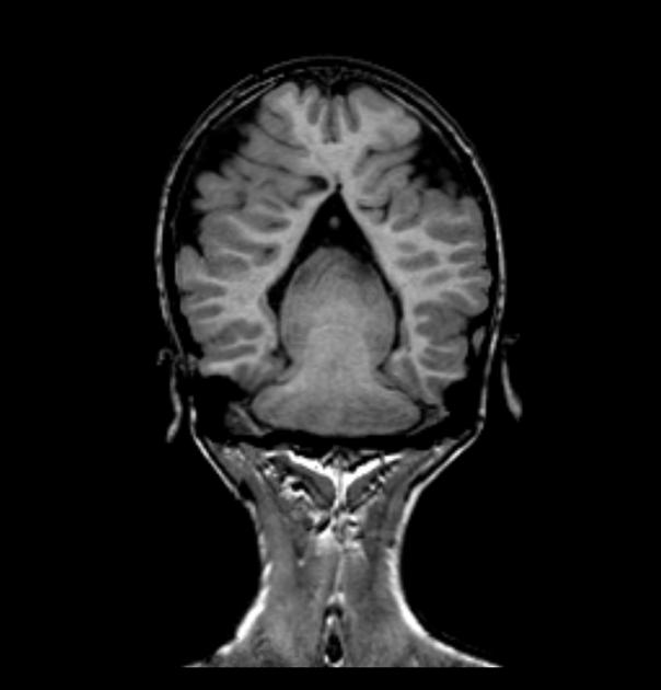
vertically orientated Sylvian fissures which are abnormally connected across the midline over the vertex of the brain
dorsal cyst
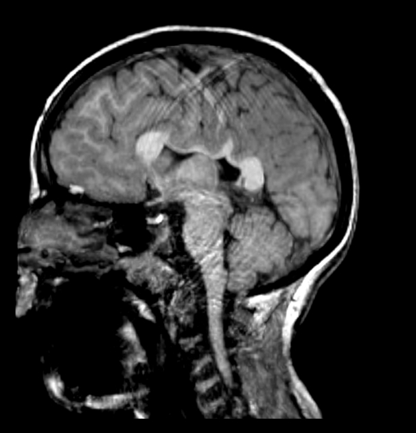
hypoplasia or aplasia of the body of corpus callosum
presence of an interhemispheric fissure
absent septum pellucidum, similar to the other forms of holoprosencephaly spectrum
separate frontal and occipital lobes
olfactory lobes are present
Treatment and prognosis
Unfortunately, as is the case with most congenital structural brain abnormalities, no specific treatment is available. The degree of neurological impairment is variable.
History and etymology
This entity was described by Barkovich and Quint in 1993.
Differential diagnosis
-
usually affects the anterior and inferior parts of the prosencephalon
more frequently associated with a dorsal cyst
cerebellar abnormalities are less common 1
-
the coronal orientation of the Sylvian fissures may mimic bilateral schizencephaly, but no extension to the ventricles is present


 Unable to process the form. Check for errors and try again.