
Tongue
Citation, DOI, disclosures and article data
At the time the article was created Henry Knipe had no recorded disclosures.
View Henry Knipe's current disclosuresAt the time the article was last revised Craig Hacking had the following disclosures:
- Philips Australia, Paid speaker at Philips Spectral CT events (ongoing)
These were assessed during peer review and were determined to not be relevant to the changes that were made.
View Craig Hacking's current disclosures- Tongues
- Frenulum of the tongue
- Frenula of the tongue
The tongue is a complex, principally muscular structure that extends from the oral cavity to the oropharynx. It has important roles in speech, swallowing and taste.
On this page:
Gross anatomy
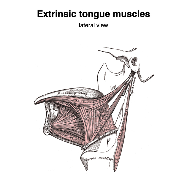
The tongue has a tip, dorsum, inferior surface and root. The tongue is made of a midline lingual septum and hyoglossus membrane, and multiple muscles 1,2,4. The muscles are divided into intrinsic and extrinsic muscle groups:
-
intrinsic muscles of the tongue which do not have attachments outside the tongue and whose action is to alter the shape of the tongue:
-
extrinsic muscles of the tongue (mnemonic) which have attachments outside the tongue and therefore their actions alter the position of the tongue:
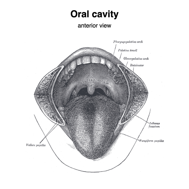
The tongue is divided into two parts at the level of the circumvallate papillae 1,3:
-
mobile tongue: anterior two-thirds; part of the oral cavity
includes root of tongue, which is considered separately due to its importance in oropharyngeal cancer
base of tongue: posterior one-third; fixed; part of the oropharynx
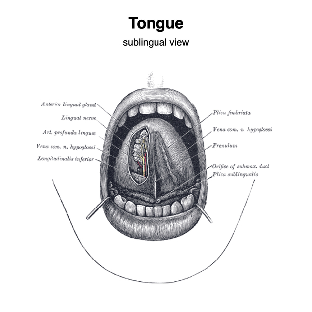
The tongue is covered by a mucosa, which is roughened on the dorsal surface covered by filiform, fungiform and circumvallate papillae. Posteriorly, the base of the tongue contains the lingual tonsils 4.
On its inferior surface the tongue is usually joined to the floor of the mouth by a thin midline membrane, the frenulum of the tongue.
ADVERTISEMENT: Supporters see fewer/no ads
Arterial supply
lingual artery (principally) but also branches from the facial and ascending pharyngeal arteries 4
Venous drainage
follows arterial supply draining to the lingual, facial and/or internal jugular veins 4
Innervation
hypoglossal nerve (CN XII): intrinsic and extrinsic muscles (except palatoglossus muscle, which is supplied by the pharyngeal plexus)
-
sensory supply to the anterior two-thirds
special sensory (taste) fibers diverge from the lingual nerve and travel with the facial nerve (CN VII) via chorda tympani
glossopharyngeal nerve (CN IX): sensory supply to posterior one-third 2
Lymphatic drainage
The dorsal mucosal surface of the pharyngeal part of the tongue contains groups of lymphoid follicles forming the lingual tonsils.
The anterior tongue drains to several nodal groups:
apex: drains to submental and submandibular nodes
body: drains to submandibular nodes then to the deep cervical nodes (especially the jugulodigastric and juguloomohyoid nodes)
The posterior tongue drains directly to deep cervical nodes.
Of clinical significance in tumors approaching the midline, central regions of the tongue may drain bilaterally, especially if lymphatic vessels on one side are obstructed.
ADVERTISEMENT: Supporters see fewer/no ads
History and etymology
The word tongue is derived from an Old English word tunge, meaning the organ of phonation, or speech itself, and ultimately is thought to be derived from lingua, the Latin for tongue 7.
Related pathology
-
neoplastic
hemangioma
lipoma
-
non neoplastic
References
- 1. Fang WS, Wiggins RH, Illner A et-al. Primary lesions of the root of the tongue. Radiographics. 2011;31 (7): 1907-22. doi:10.1148/rg.317095738 - Pubmed citation
- 2. Head and Neck Imaging, An Issue of Radiologic Clinics of North America. Elsevier Health Sciences. (2014) ISBN:0323342027. Read it at Google Books - Find it at Amazon
- 3. Law CP, Chandra RV, Hoang JK et-al. Imaging the oral cavity: key concepts for the radiologist. Br J Radiol. 2011;84 (1006): 944-57. Br J Radiol (full text) - doi:10.1259/bjr/70520972 - Free text at pubmed - Pubmed citation
- 4. Clinical Head and Neck Anatomy for Surgeons. CRC Press. ISBN:144415737X. Read it at Google Books - Find it at Amazon
- 5. Mcminn. Last's Anatomy. ISBN: 9780729537520
- 6. Keith L. Moore, Arthur F. Dalley. Clinically Oriented Anatomy. ISBN: 9780683061413
- 7. Robert K. Barnhart, Sol Steinmetz. Chambers Dictionary of Etymology. (1999) ISBN: 9780550142306
- 8. Susan Standring. Gray's Anatomy. ISBN: 9780702052309 https://drive.google.com/file/d/0BwQj92tZP0i3bEk5YmR0LUI1bFE/view?usp=sharing
- 9. Robert H. Whitaker, Neil R. Borley. Instant Anatomy. ISBN: 9780632054039
Incoming Links
- Pyogenic granuloma
- Genioglossus muscle
- Ludwig angina
- Salivary glands
- Thyroglossal duct
- Neck-tongue syndrome
- Squamous cell carcinoma (oral cavity)
- Organomegaly
- Intrinsic muscles of the tongue
- Hypoglossal nerve
- Squamous cell carcinoma (tongue)
- Superior longitudinal muscle of the tongue
- Submandibular gland
- Synovial sarcoma
- Submental artery
- Oropharynx
- Submandibular duct
- Bright tongue sign
- Pharynx
- Lingula (disambiguation)
- Extrinsic tongue muscles (Gray's illustration)
- Tongue and oral cavity (Gray's illustrations)
- Intramuscular haemangioma (tongue)
- Tongue vessels (Gray's illustration)
- Lymphatics of the tongue (Gray's illustration)
- Submandibular gland haemangioma
- Squamous cell carcinoma of the tongue
- Ludwig angina
- Glossopharyngeal nerve schwannoma
Related articles: Anatomy: Head and neck
- skeleton of the head and neck
-
cranial vault
- scalp (mnemonic)
- fontanelle
-
sutures
- calvarial
- facial
- frontozygomatic suture
- frontomaxillary suture
- frontolacrimal suture
- frontonasal suture
- temporozygomatic suture
- zygomaticomaxillary suture
- parietotemporal suture (parietomastoid suture)
- occipitotemporal suture (occipitomastoid suture)
- sphenofrontal suture
- sphenozygomatic suture
- spheno-occipital suture (not a true suture)
- lacrimomaxillary suture
- nasomaxillary suture
- internasal suture
- basal/internal
- skull landmarks
- frontal bone
- temporal bone
- parietal bone
- occipital bone
- skull base (foramina)
-
facial bones
- midline single bones
- paired bilateral bones
- cervical spine
- hyoid bone
- laryngeal cartilages
-
cranial vault
- muscles of the head and neck
- muscles of the tongue (mnemonic)
- muscles of mastication
-
facial muscles
- epicranius muscle
- circumorbital and palpebral muscles
- nasal muscles
-
buccolabial muscles
- elevators, retractors and evertors of the upper lip
- levator labii superioris alaeque nasalis muscle
- levator labii superioris muscle
- zygomaticus major muscle
- zygomaticus minor muscle
- levator anguli oris muscle
- malaris muscle
- risorius muscle
- depressors, retractors and evertors of the lower lip
- depressor labii inferioris muscle
- depressor anguli oris muscle
- mentalis muscle
- compound sphincter
-
orbicularis oris muscle
- incisivus labii superioris muscle
- incisivus labii inferioris muscle
-
orbicularis oris muscle
- muscle of mastication
- modiolus
- elevators, retractors and evertors of the upper lip
- muscles of the middle ear
- orbital muscles
- muscles of the soft palate
- pharyngeal muscles
- suprahyoid muscles
- infrahyoid muscles
- intrinsic muscles of the larynx
- muscles of the neck
- platysma muscle
- longus colli muscle
- longus capitis muscle
- scalenus anterior muscle
- scalenus medius muscle
- scalenus posterior muscle
- scalenus pleuralis muscle
- sternocleidomastoid muscle
-
suboccipital muscles
- rectus capitis posterior major muscle
- rectus capitis posterior minor muscle
- obliquus capitis superior muscle
- obliquus capitis inferior muscle
- accessory muscles of the neck
- deep cervical fascia
-
deep spaces of the neck
- anterior cervical space
- buccal space
- carotid space
- danger space
- deep cervical fascia
- infratemporal fossa
- masticator space
- parapharyngeal space
- stylomandibular tunnel
- parotid space
- pharyngeal (superficial) mucosal space
- perivertebral space
- posterior cervical space
- pterygopalatine fossa
- retropharyngeal space
- suprasternal space (of Burns)
- visceral space
- surgical triangles of the neck
- orbit
- ear
- paranasal sinuses
- upper respiratory tract
- viscera of the neck
- blood supply of the head and neck
-
arterial supply
-
common carotid artery
- carotid body
- carotid bifurcation
- subclavian artery
- variants
-
common carotid artery
- venous drainage
-
arterial supply
- innervation of the head and neck
-
cranial nerves
- olfactory nerve (CN I)
- optic nerve (CN II)
- oculomotor nerve (CN III)
- trochlear nerve (CN IV)
-
trigeminal nerve (CN V) (mnemonic)
- trigeminal ganglion
- ophthalmic division
- maxillary division
- mandibular division
- abducens nerve (CN VI)
- facial nerve (CN VII)
-
vestibulocochlear nerve (CN VIII)
- vestibular ganglion (Scarpa's ganglion)
- glossopharyngeal nerve (CN IX)
- vagus nerve (CN X)
- (spinal) accessory nerve (CN XI)
- hypoglossal nerve (CN XII)
- parasympathetic ganglia of the head and neck
- cervical sympathetic ganglia
- greater occipital nerve
- third occipital nerve
-
cervical plexus
- muscular branches
- longus capitis
- longus colli
- scalenes
- geniohyoid
- thyrohyoid
-
ansa cervicalis
- omohyoid (superior and inferior bellies separately)
- sternothyroid
- sternohyoid
- phrenic nerve
- contribution to the accessory nerve (CN XI)
- cutaneous branches
- muscular branches
- brachial plexus
- pharyngeal plexus
-
cranial nerves
- lymphatic drainage of the head and neck
- embryological development of the head and neck


 Unable to process the form. Check for errors and try again.