
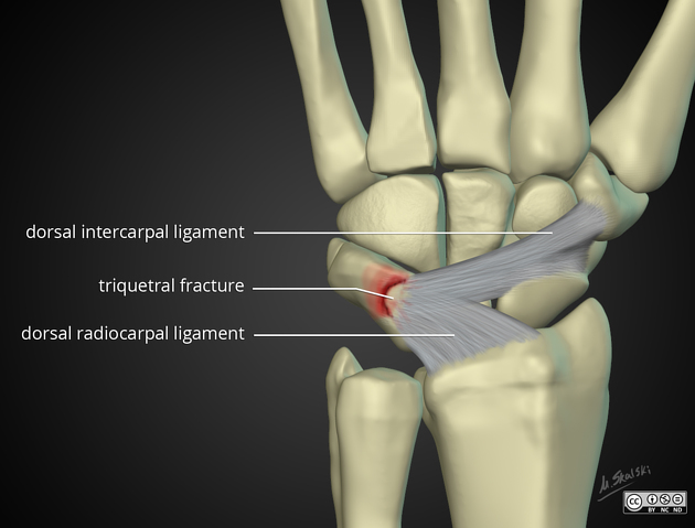
Triquetral fracture
Citation, DOI, disclosures and article data
At the time the article was created The Radswiki had no recorded disclosures.
View The Radswiki's current disclosuresAt the time the article was last revised Martin Bundi Rugendo had no financial relationships to ineligible companies to disclose.
View Martin Bundi Rugendo's current disclosures- Triquetrum fracture
- Triquetral fractures
Triquetral fractures are carpal bone fractures generally occurring on the dorsal surface of the triquetrum. The triquetral may be fractured by means of impingement from the ulnar styloid, shear forces, or avulsion from strong ligamentous attachments. They are the second commonest carpal bone fracture, after the scaphoid.
On this page:
Clinical presentation
Commonest history is trauma to the outstretched hand with carpal extension 4:
- pain is usually on the ulnar aspect of the wrist, exacerbated by extension/flexion of the wrist
- swelling over the dorsum of the hand with a tender dorsal aspect of triquetrum may be found on exam
Pathology
The usual injury mechanism is falling onto an outstretched hand in ulnar deviation. Less commonly, it may be caused by a direct blow to the dorsum of the hand, a situation where commonly other carpal fractures are seen.
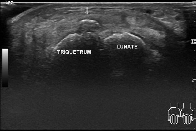
Radiographic features
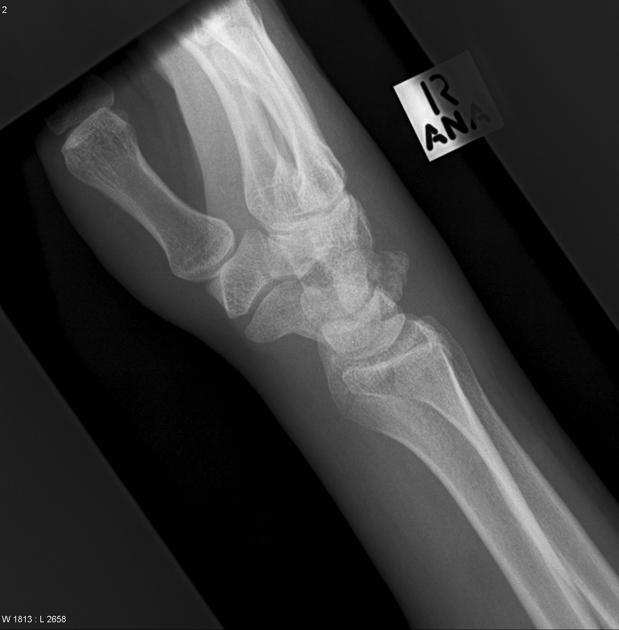
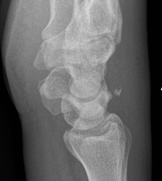
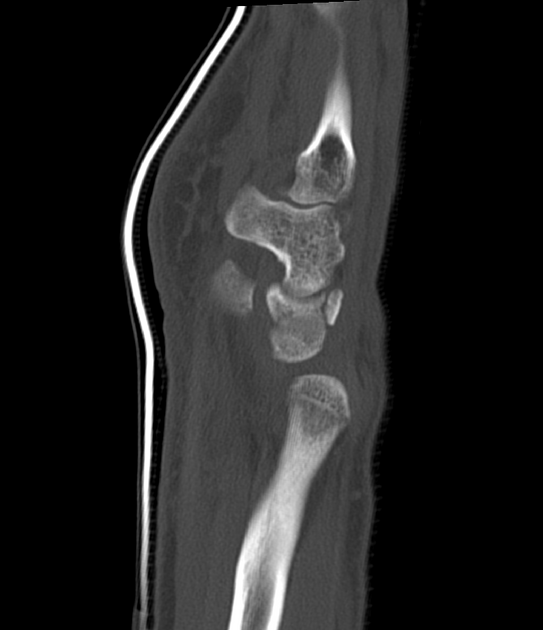
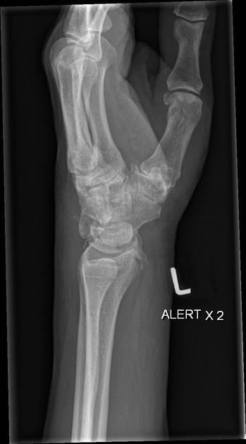
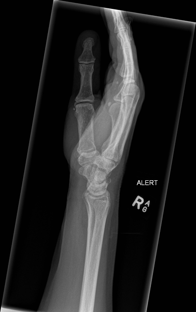
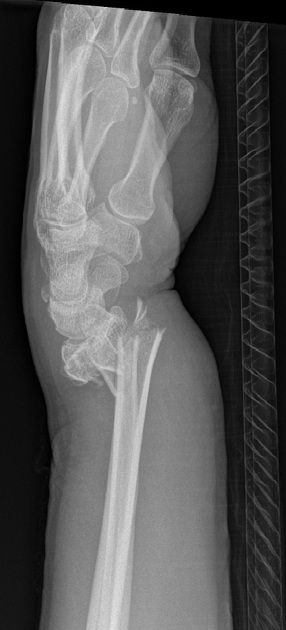
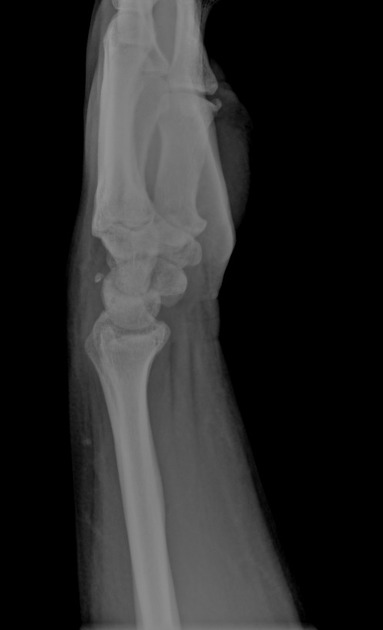
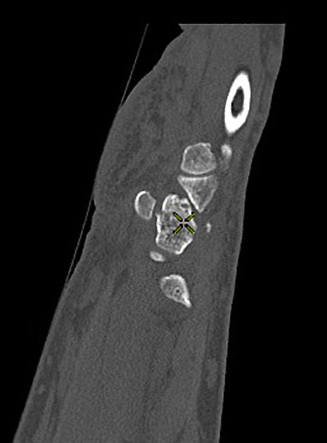
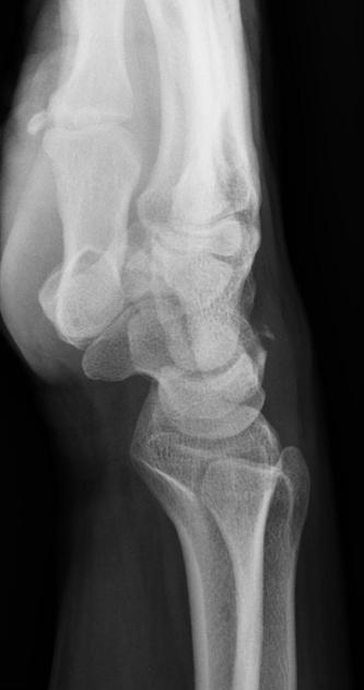
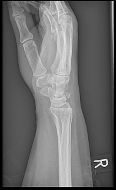
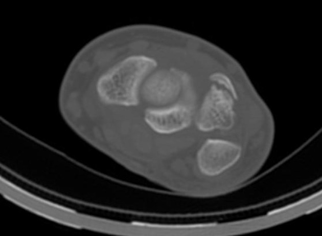
There are three fracture patterns often observed, dorsal avulsion fractures, triquetral body fractures and volar avulsion fractures 3. Dorsal avulsion fractures account for about 95% all triquetral fractures, most of the remainder are body fractures 4.
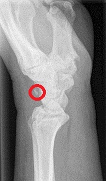
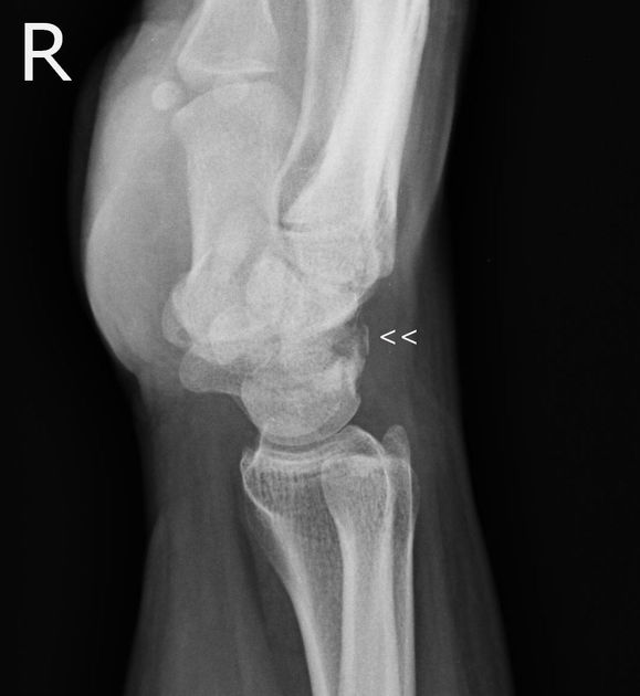
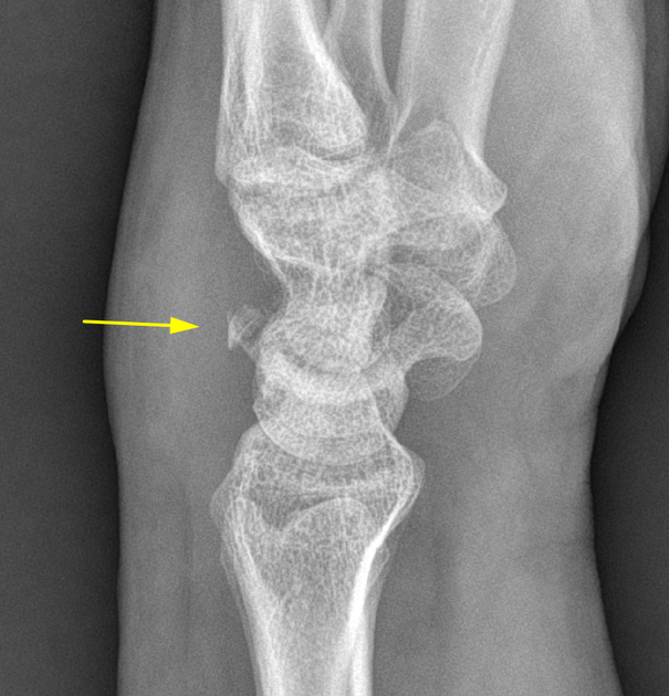
Dorsal avulsion fracture
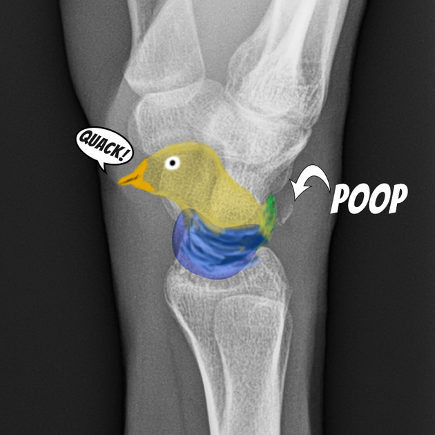
On plain film, dorsal avulsion injuries are best detected on a lateral projection, where typically an avulsed flake of bone is identified lying posteriorly to the triquetral bone (see pooping duck sign). CT or MR may be more sensitive than conventional radiographs for detection of avulsion injuries.
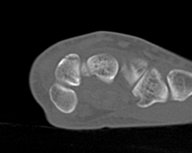
Triquetral body fracture
Triquetral body fractures appear as clear fracture lines through the body, they are best seen on the oblique projection of the wrist, although cross sectional imaging may be required to further elucidate extent.
Volar avulsion fractures
Volar avulsion fractures are avulsions of the palmar ulnar triquetral/lunotriquetral ligament and are best seen on a radial deviation projection of the wrist 3.
Treatment and prognosis
Surgical intervention is rarely required, but a persistently symptomatic chip fracture may require excision.
Differential diagnosis
Imaging differential considerations include:
References
- 1. Goldfarb C, Yin Y, Gilula L, Fisher A, Boyer M. Wrist Fractures: What the Clinician Wants to Know. Radiology. 2001;219(1):11-28. doi:10.1148/radiology.219.1.r01ap1311 - Pubmed
- 2. Becce F, Theumann N, Bollmann C et al. Dorsal Fractures of the Triquetrum: MRI Findings with an Emphasis on Dorsal Carpal Ligament Injuries. AJR Am J Roentgenol. 2013;200(3):608-17. doi:10.2214/AJR.12.8736 - Pubmed
- 3. Suh N, Ek E, Wolfe S. Carpal Fractures. J Hand Surg Am. 2014;39(4):785-91; quiz 791. doi:10.1016/j.jhsa.2013.10.030 - Pubmed
- 4. Guo R, Cardenas J, Wu C. Triquetral Fractures Overview. Curr Rev Musculoskelet Med. 2021;14(2):101-6. doi:10.1007/s12178-021-09692-w - Pubmed
Incoming Links
- Pooping duck sign
- Animal and animal produce inspired signs
- Fall onto an outstretched hand
- Wrist radiograph (checklist)
- Final FRCR Part B rapid reporting
- Mayfield classification of carpal instability (perilunate instability)
- Carpal bone fractures
- Hand radiograph (checklist)
- Wrist radiograph (an approach)
- Triquetrum
- Upper extremity fractures
- Isolated capitate-trapezoid coalition
- Triquetral fracture - pooping duck sign
- Triquetral fracture - pooping duck sign
- Proximal carpal row fracture-dislocation
- Pooping duck sign
- Triquetral fracture - pooping duck sign
- Triquetral fracture - pooping duck sign
- Triquetral fracture-pooping duck sign
- Bilateral Colles fractures
- Triquetral fracture
- Triquetral fracture and central perforation of the triangular fibrocartilage
- Triquetral bone fracture
- Isolated triquetral body fracture
- Fractured distal radius, distal ulna and triquetrum
- Triquetral fracture - pooping duck sign
- Distal radius fracture with triquetral avulsion
- Triquetral fracture
- Triquetral fracture
- Triquetral fracture
- Triquetral fracture
Related articles: Fractures
-
fracture
- terminology
- fracture location
- diaphyseal fracture
- metaphyseal fracture
- physeal fracture
- epiphyseal fracture
- fracture types
- avulsion fracture
- articular surface injuries
- complete fracture
- incomplete fracture
- infraction
- compound fracture
- pathological fracture
- stress fracture
- fracture displacement
- fracture location
- fracture healing
- skull fractures
-
facial fractures
- fractures involving a single facial buttress
- alveolar process fractures
- frontal sinus fracture
- isolated zygomatic arch fractures
- mandibular fracture
- nasal bone fracture
- orbital blow-out fracture
- paranasal sinus fractures
- complex fractures
- dental fractures
- fractures involving a single facial buttress
-
spinal fractures
- classification (AO Spine classification systems)
-
cervical spine fracture classification systems
- AO classification of upper cervical injuries
- AO classification of subaxial injuries
- Anderson and D'Alonzo classification (odontoid fracture)
- Roy-Camille classification (odontoid process fracture)
- Gehweiler classifcation (atlas fractures)
- Levine and Edwards classification (hangman fracture)
- Allen and Ferguson classification (subaxial spine injuries)
- subaxial cervical spine injury classification (SLIC)
- thoracolumbar spinal fracture classification systems
- three column concept of spinal fractures (Denis classification)
- classification of sacral fractures
-
cervical spine fracture classification systems
- spinal fractures by region
- spinal fracture types
- classification (AO Spine classification systems)
- rib fractures
- sternal fractures
-
upper limb fractures
- classification
- Rockwood classification (acromioclavicular joint injury)
- AO classification (clavicle fracture)
- Neer classification (clavicle fracture)
- Neer classification (proximal humeral fracture)
- AO classification (proximal humeral fracture)
- AO/OTA classification of distal humeral fractures
- Milch classification (lateral humeral condyle fracture)
- Weiss classification (lateral humeral condyle fracture)
- Bado classification of Monteggia fracture-dislocations (radius-ulna)
- Mason classification (radial head fracture)
- Frykman classification (distal radial fracture)
- Mayo classification (scaphoid fracture)
- Hintermann classification (gamekeeper's thumb)
- Eaton classification (volar plate avulsion injury)
- Keifhaber-Stern classification (volar plate avulsion injury)
- upper limb fractures by region
- shoulder
- clavicular fracture
-
scapular fracture
- acromion fracture
- coracoid process fracture
- glenoid fracture
- humeral head fracture
- proximal humeral fracture
- humeral neck fracture
- arm
- elbow
- forearm
- wrist
-
carpal bones
- scaphoid fracture
- lunate fracture
- capitate fracture
- triquetral fracture
- pisiform fracture
- hamate fracture
- trapezoid fracture
- trapezium fracture
- hand
- shoulder
- classification
- lower limb fractures
- classification by region
- pelvic fractures
- hip fractures
- Pipkin classification (femoral head fracture)
- Garden classification (hip fracture)
- American Academy of Orthopaedic Surgeons classification (periprosthetic hip fracture)
- Cooke and Newman classification (periprosthetic hip fracture)
- Johansson classification (periprosthetic hip fracture)
- Vancouver classification (periprosthetic hip fracture)
- femoral
- knee
- Schatzker classification (tibial plateau fracture)
- AO classification of distal femur fractures
- Meyers and McKeevers classification (anterior cruciate ligament avulsion fracture)
- tibia/fibula
- Watson-Jones classification (tibial tuberosity avulsion fracture)
- ankle
- foot
- Berndt and Harty classification (osteochondral lesions of the talus)
- Sanders CT classification (calcaneal fracture)
- Hawkins classification (talar neck fracture)
- Myerson classification (Lisfranc injury)
- Nunley-Vertullo classification (Lisfranc injury)
- pelvis and lower limb fractures by region
- pelvic fracture
- sacral fracture
- coccygeal fracture
-
hip
- acetabular fracture
- femoral head fracture
-
femoral neck fracture
- subcapital fracture
- transcervical fracture
- basicervical fracture
-
trochanteric fracture
- pertrochanteric fracture
- intertrochanteric fracture
- subtrochanteric fracture
- femur
- mid-shaft fracture
- bisphosphonate-related fracture
- distal femoral fracture
- knee
- avulsion fractures
- Segond fracture
- reverse Segond fracture
- anterior cruciate ligament avulsion fracture
- posterior cruciate ligament avulsion fracture
- arcuate complex avulsion fracture (arcuate sign)
- biceps femoris avulsion fracture
- iliotibial band avulsion fracture
- semimembranosus tendon avulsion fracture
- Stieda fracture (MCL avulsion fracture)
- patellar fracture
- tibial plateau fracture
- avulsion fractures
- leg
- tibial tuberosity avulsion fracture
- tibial shaft fracture
- fibular shaft fracture
- Maisonneuve fracture
- ankle
- foot
- tarsal bones
- metatarsal bones
- phalanges
- classification by region
- terminology
Related articles: Wrist pathology
- alignment
- wrist fractures and dislocations
- distal radial fracture
- paediatric
- carpal bones
- Mayfield classification of carpal instability
- carpal instability
- osteonecrosis
- triangular fibrocartilaginous complex (TFCC) injuries
- ulnar-sided wrist impaction and impingement syndromes
- soft tissue and tendons
- arthritides


 Unable to process the form. Check for errors and try again.