
Urinomas, or uriniferous fluid collections, are urine collections usually found in the retroperitoneum, most commonly in the perirenal space, as a consequence of renal tract leakage caused by urinary obstruction, trauma, or post-instrumentation.
On this page:
Terminology
As there is no definitive distinction between the terms urinoma and urine leak in the literature, the terms are often used interchangeably. This article will treat them differently with the former as a consequence of the latter, i.e. urinomas are encapsulated urine collections due to urine leakage.
Pathology
As urine extravasates into the retroperitoneum, it can cause lipolysis of the surrounding fat with resultant encapsulation of urine, forming a urinoma.
An elevated ascitic fluid creatinine to serum creatinine ratio over 1.18 identifies the fluid with urinary origin, with a sensitivity and specificity of 78% and 88% respectively based on a retrospective study 5.
Urinomas are most often due to urinary obstruction secondary to 2,3:
renal pelvis, ureteric, or bladder cancer
various conditions causing bladder outlet obstruction
Urinomas also may be due to:
abdominopelvic trauma, e.g. renal trauma or bladder rupture
surgery, e.g. ureteric injury, ureteroileal anastomotic leak
diagnostic instrumentation, e.g. ESWL, ureteroscopy
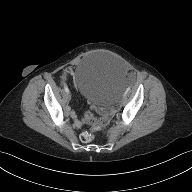
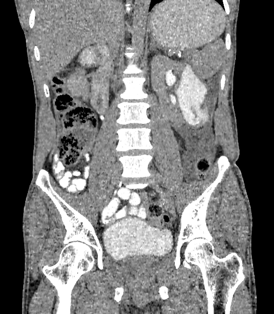
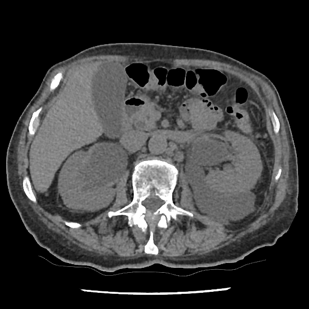
Location and morphology
cystic mass in perirenal space: localized perirenal urinoma (most common)
cystic mass filling entire perirenal space: diffuse perirenal urinoma
sickle-shaped collection: subcapsular urinoma
encapsulated expanding intrarenal cystic mass separating renal tissue fragments: intrarenal urinoma 3
Radiographic features
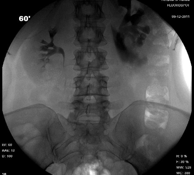
Fluoroscopy
Intravenous pyelogram
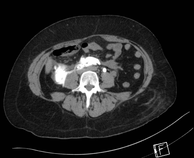
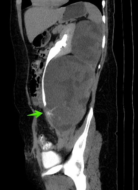
Contrast extravasates outside of the collecting system, into the surrounding retroperitoneal tissues.
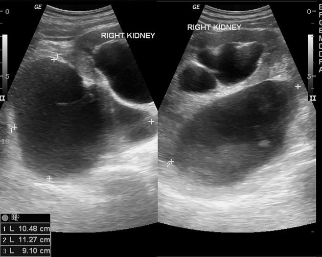
Ultrasound
Presents as a thinned-walled anechoic collection usually found partially contouring any portion of the renal tracts.
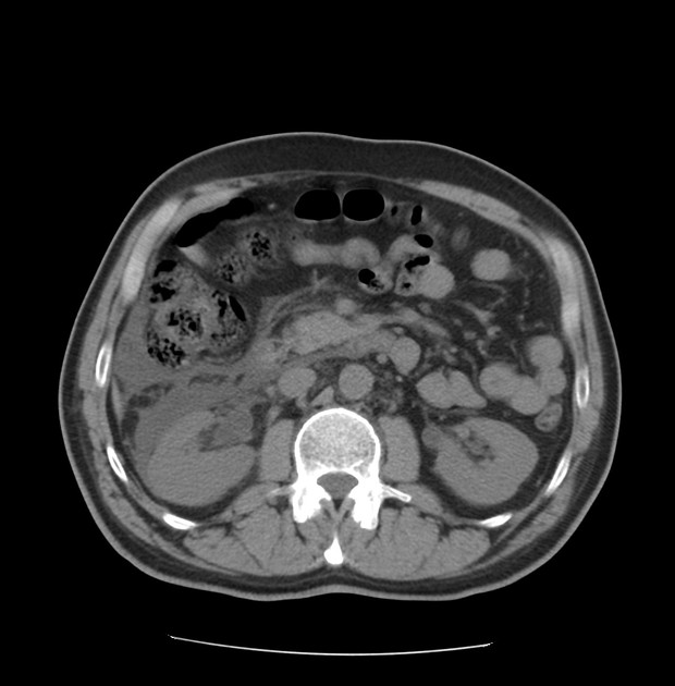
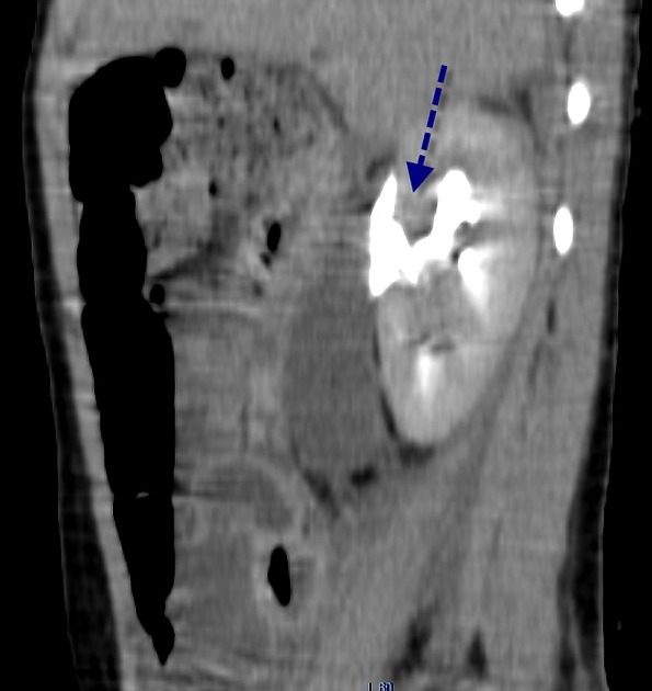
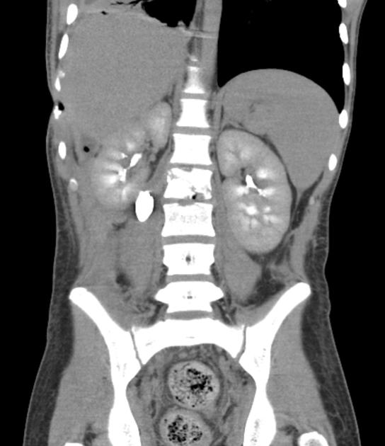
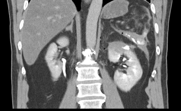
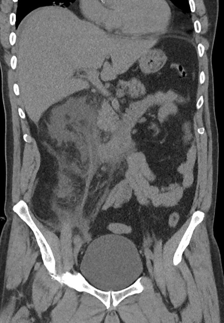
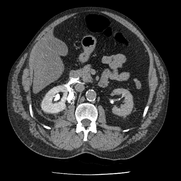
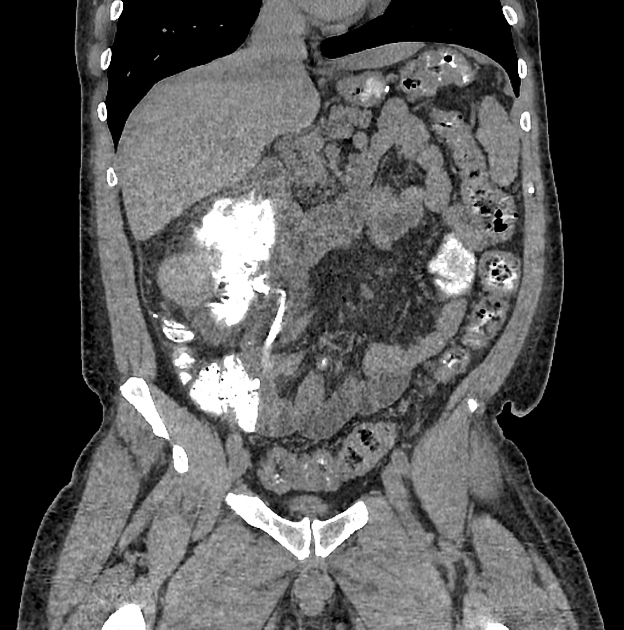
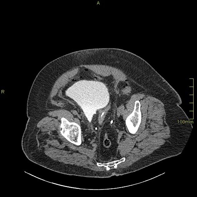
CT and MRI
On CT and MRI, a urinoma shows water attenuation, low signal intensity on T1-weighted imaging, and very high signal intensity on T2-weighted imaging, similar to simple fluid elsewhere in the body.
Urine leakage is usually directly demonstrated on contrast-enhanced studies on the excretory phase due to direct contrast extravasation from the urinary tract 2.
Treatment and prognosis
small urinomas are usually treated conservatively
larger, persistent or symptomatic urinomas: percutaneous drainage is often used with the treatment of the underlying cause of urine leakage


 Unable to process the form. Check for errors and try again.