
Presentation
The child was involved in a motor vehicle accident and received blunt abdominal trauma.
Patient Data
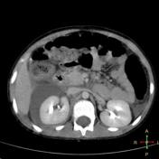

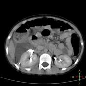

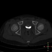

Axial portovenous phase images reveal a large large fluid collection in the right anterior perirenal space and the hepatorenal space. Small amount of fludi in the right paracolic gutter and a small right pleural effusion.
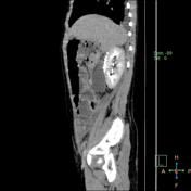
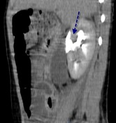
Delayed images reveal a connection between this fluid and the upper calyx. The collection filled gradually with contrast, indicating a urine leak.
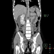
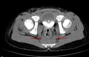
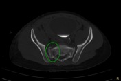
The pelvic images show a pre-sacral hematoma and bone window images show a sagittaly oriented right sacral ala fracture with small detached fragment.
Diagnosis: Post traumatic urinoma
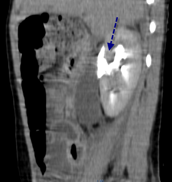
Illustrated selected images from the study, with annotations to illustrate key findings:
- dashed blue arrow refers to the communication between upper calyx and the urinoma
- solid red arrows refer to the presacral hematoma
- green circle shows the fracture right ala with a small bony fragment
Case Discussion
An extra-renal urine collection (urinoma) can occur following acute urinary tract obstruction, trauma or iatrogenic after surgery or renal intervention e.g. biopsy.
A urine leak usually collects beneath the renal capsule (sub-capsular) or outside the capsule in the perirenal space.
Contrast study e.g. IVU or C-IVU are the main diagnostic methods.


 Unable to process the form. Check for errors and try again.