
White matter tracts
Last revised by Tariq Walizai
on 9 Jul 2024
Citation, DOI, disclosures and article data
Citation:
Abdrabou A, Walizai T, Al Kabbani A, et al. White matter tracts. Reference article, Radiopaedia.org (Accessed on 21 Mar 2025) https://doi.org/10.53347/rID-22721
rID:
22721
Article created:
19 Apr 2013,
Ahmed Abdrabou
Disclosures:
At the time the article was created Ahmed Abdrabou had no recorded disclosures.
View Ahmed Abdrabou's current disclosures
Last revised:
9 Jul 2024,
Tariq Walizai
Disclosures:
At the time the article was last revised Tariq Walizai had no financial relationships to ineligible companies to disclose.
View Tariq Walizai's current disclosures
Revisions:
26 times, by
15 contributors -
see full revision history and disclosures
Systems:
Sections:
Tags:
Synonyms:
- White matter fibres
- White matter fibres
- White matter bundles
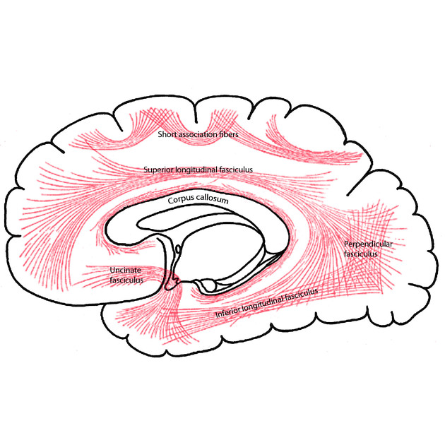
White matter tracts in the brain, also known as white matter fibres, are classified into three categories:
The white matter can be observed well on T1W, T2W and FLAIR sequences. Some white matter tracts are quite well demonstrated especially on T2W and FLAIR sequences because of their specific location and due to differences in the myelination and water content seen in the paediatric population. However, diffusion tensor imaging (DTI) provides more detailed and clinically beneficial information.
References
- 1. A. R. Crossman, David Neary. Neuroanatomy. (2000), Ch (13) 146-149, 2000. ISBN: 9780443062162
- 2. Jellison BJ, Field AS, Medow J, Lazar M, Salama MS, and Alexander AL. Diffusion Tensor Imaging of Cerebral White Matter: A Pictorial Review of Physics, Fiber Tract Anatomy, and Tumor Imaging Patterns. American Journal of Neuroradiology 2004; 25:356-369.
- 3. Kenichi Oishi, Andreia V. Faria, Peter C. M. van Zijl, Susumu Mori. MRI Atlas of Human White Matter. (2010) ISBN: 9780123820815
- 4. Wakana S, Jiang H, Nagae-Poetscher LM, van Zijl PC, Mori S. Fiber tract-based atlas of human white matter anatomy. (2004) Radiology. 230 (1): 77-87. doi:10.1148/radiol.2301021640 - Pubmed
- 5. Catani M, Mesulam M. The arcuate fasciculus and the disconnection theme in language and aphasia: history and current state. (2008) Cortex; a journal devoted to the study of the nervous system and behavior. 44 (8): 953-61. doi:10.1016/j.cortex.2008.04.002 - Pubmed
- 6. Tremblay P, Dick AS. Broca and Wernicke are dead, or moving past the classic model of language neurobiology. (2016) Brain and language. 162: 60-71. doi:10.1016/j.bandl.2016.08.004 - Pubmed
- 7. Wycoco V, Shroff M, Sudhakar S, Lee W. White matter anatomy: what the radiologist needs to know. (2013) Neuroimaging clinics of North America. 23 (2): 197-216. doi:10.1016/j.nic.2012.12.002 - Pubmed
Incoming Links
Articles:
- Association fibres of the brain
- Commissural fibres of the brain
- Corpus callosum
- Corona radiata
- External capsule
- Extra-axial
- Telencephalon
- Diffusion tensor imaging and fiber tractography
- Anterior commissure
- White matter tracts
- Extreme capsule
- White matter
- Central nervous system embryology
- Medical abbreviations and acronyms (W)
- Projection fibres of the brain
- Dorsolateral fasciculus
- Inferior cerebellar peduncle
Cases:
- MRI-Tractography for detecting the position of corticospinal tract in preoperative meningioma
- Thalamic radiations in vivo using diffusion tensor imaging (DTI)
- Agenesis of the corpus callosum - 3D reconstruction of Probst bundles
- Association fibres (Gray's illustration)
- Association fibres (Gray's illustration)
Related articles: Anatomy: Brain
-
brain
- grey matter
- white matter
-
cerebrum
-
cerebral hemisphere (telencephalon)
- cerebral lobes and gyri
- frontal lobe
- parietal lobe
-
occipital lobe
- occipital pole
- lingual gyrus
- fusiform gyrus (Brodmann area 37)
- calcarine (visual) cortex
- cuneus
- temporal lobe
- basal forebrain
- limbic system
- insula
-
cerebral sulci and fissures (A-Z)
- calcarine fissure
- callosal sulcus
- central (Rolandic) sulcus
- cingulate sulcus
- collateral sulcus
- inferior frontal sulcus
- inferior occipital sulcus
- inferior temporal sulcus
- interhemispheric fissure
- intraparietal sulcus
- lateral (Sylvian) sulcus
- lateral occipital sulcus
- marginal sulcus
- occipitotemporal sulcus
- olfactory sulcus
- paracentral sulcus
- paraolfactory sulcus
- parieto-occipital fissure
- posterior parolfactory sulcus
- precentral sulcus
- preoccipital notch
- postcentral sulcus
- rhinal sulcus
- rostral sulcus
- subparietal sulcus
- superior frontal sulcus
- superior occipital sulcus
- superior temporal sulcus
- cortical histology
- cerebral lobes and gyri
- white matter tracts
- deep grey matter
-
pituitary gland
- posterior pituitary and stalk (part of diencephalon)
- anterior pituitary
- inferior hypophyseal arterial circle
- diencephalon
-
cerebral hemisphere (telencephalon)
-
brainstem
- midbrain (mesencephalon)
- pons (part of metencephalon)
- medulla oblongata (myelencephalon)
- white matter
-
grey matter
- non-cranial nerve
-
cranial nerve nuclei
- oculomotor nucleus
- Edinger-Westphal nucleus
- trochlear nucleus
- motor nucleus of CN V
- mesencephalic nucleus of CN V
- main sensory nucleus of CN V
- spinal nucleus of CN V
- abducent nucleus
- facial nucleus
- superior salivatory nucleus
- cochlear nuclei
- vestibular nuclei
- inferior salivatory nucleus
- solitary tract nucleus
- ambiguus nucleus
- dorsal vagal motor nucleus
- hypoglossal nucleus
-
cerebellum (part of metencephalon)
- vermis
- cerebellar hemisphere
- cerebellar peduncles
- cranial meninges (meninx primitiva)
- CSF spaces
-
cranial nerves (mnemonic)
- olfactory nerve (CN I)
- optic nerve (CN II)
- oculomotor nerve (CN III)
- trochlear nerve (CN IV)
- trigeminal nerve (CN V) (mnemonic)
- abducens nerve (CN VI)
- facial nerve (CN VII) (segments mnemonic | branches mnemonic)
-
vestibulocochlear nerve (CN VIII)
- vestibular ganglion (Scarpa's ganglion)
- glossopharyngeal nerve (CN IX)
- vagus nerve (CN X)
- spinal accessory nerve (CN XI)
- hypoglossal nerve (CN XII)
- functional neuroanatomy
- CNS development
- cerebral vascular supply
- arteries
- vascular territories
-
circle of Willis
- internal carotid artery (ICA) (segments)
- vertebral artery
-
normal variants
- intracranial arterial fenestration
- internal carotid artery (ICA)
- anterior cerebral artery (ACA)
- middle cerebral artery (MCA)
- posterior cerebral artery (PCA)
- basilar artery
- persistent carotid-vertebrobasilar artery anastomoses (mnemonic)
- vertebral artery
- ophthalmic artery
-
cerebral venous system
-
dural venous sinuses
- basilar venous plexus
- cavernous sinus (mnemonic)
- clival diploic veins
- inferior petro-occipital vein
- inferior petrosal sinus
- inferior sagittal sinus
- intercavernous sinus
- internal carotid artery venous plexus of Rektorzik
- jugular bulb
- marginal sinus
- occipital sinus
- sigmoid sinus
- sphenoparietal sinus
- straight sinus
- superior petrosal sinus
- superior sagittal sinus
- torcula herophili
- transverse sinus
-
cerebral veins
-
superficial veins of the brain
- superior cerebral veins (superficial cerebral veins)
- inferior cerebral veins
- superficial middle cerebral vein
- superior anastomotic vein (of Trolard)
- inferior anastomotic vein (of Labbe)
-
superficial veins of the brain
-
deep veins of the brain
- great cerebral vein (of Galen)
- venous circle of Trolard
- normal variants
-
dural venous sinuses
- arteries
- glymphatic pathway


 Unable to process the form. Check for errors and try again.