
Presentation
Work up for abdominal pain and progressive distention.
Patient Data


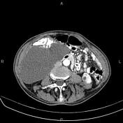
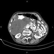
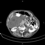
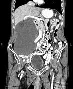

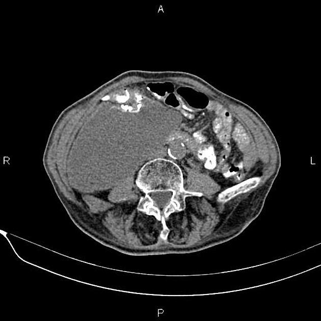
A 185×110×115 mm cystic lesion with anterior wall calcification and without enhancing solid component is seen in right abdominopelvic spaces. Several internal smaller daughter cysts are also observed.
Intra and extrahepatic bile ducts are dilated, and CBD measured 10 mm in caliber.
A few nonenhanced simple cortical cysts are seen in both kidneys, with maximum diameters of 20 mm.
Mild to moderate hydroureteronephrosis with diffuse urothelial wall thickening is seen on the left side. No renal or ureteral stones are noted bilaterally.
Diffuse but mild urinary bladder wall thickening is present.
The prostate gland is enlarged.
Degenerative changes such as osteophytosis are seen in the lumbar spine.
Evidence of DHS insertion is present at the proximal of both femurs.
Case Discussion
The patient underwent surgical resection of the sizable abdominopelvic cyst, and histopathologic findings confirmed a hydatid cyst. Retroperitoneal hydatid cyst is generally seen secondary to intraperitoneal (mainly hepatic) infection. Isolated occurrence is rare.


 Unable to process the form. Check for errors and try again.