
Presentation
History of total abdominal hysterectomy and bilateral salpingo-oophorectomy. Work up for multiple hepatic masses reported in ultrasound evaluation.
Patient Data
Age: 50 years
Gender: Female
From the case:
Hepatic haemangiomatosis


Download
Info

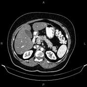
The hepatic attenuation value is less than that of the spleen, suggesting fatty liver.
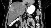
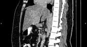
At least six masses in the liver show early peripheral nodular enhancement with centripetal filling and delayed blood pools.
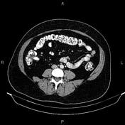
Due to prior resection, the uterus and ovaries are not seen at the anatomical location.
Degenerative changes such as osteophytosis are seen in the lumbar spine.
Grade I spondylolisthesis of L5 on S1 is present with bilateral spondylolysis.
Case Discussion
Enhancement patterns of the hepatic masses are typical for haemangiomatosis.


 Unable to process the form. Check for errors and try again.