
Presentation
Scalp swelling.
Patient Data
Age: 13 years
Note: This case has been tagged as "legacy" as it no longer meets image preparation and/or other case publication guidelines.
From the case:
Skull vault hemangioma

Download
Info

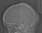
Axial CT reveals an occipital expansile intradiploic lesion with intact inner and outer tables.
From the case:
Skull vault hemangioma


Download
Info

Sagittal T1 and axial T2 MRI revealed an intraosseous occipital bone lesion with hypointense on T1 and hyperintense on T2.
Case Discussion
Diagnosis: Skull vault hemangioma


 Unable to process the form. Check for errors and try again.