
Presentation
Patient presented with diplopia.
Patient Data
Age: Middle aged
From the case:
Trigeminal schwannoma


Download
Info
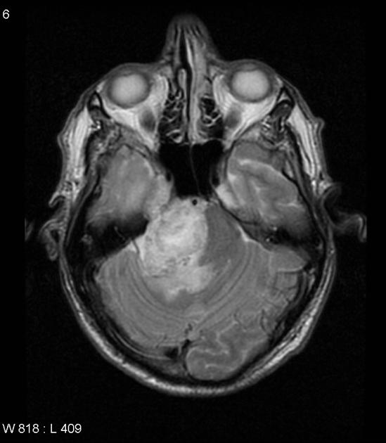
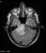
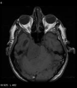
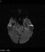
MRI through the brain demonstrates a large cerebellopontine angle mass on the right, with a component extending anteriorly into Meckle's cave. The mass is heterogenous on all sequences, and relatively high signal on T2, with avid contrast enhancement, and areas which are presumably cystic.
Download
Info
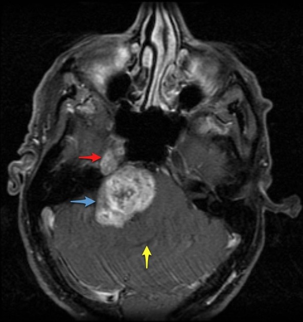
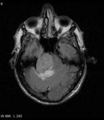
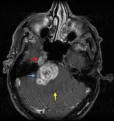
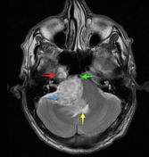
The mass has a dumbbell configuration, with a component in Meckle's cave (red arrow) and a larger one in the cerebellopontine angle (blue arrow). The fourth ventricle is markedly distorted (yellow arrow), as is the basilar artery (green arrow).
Case Discussion
This patient went on to have a craniotomy and resection of the mass. Histology confirmed a trigeminal schwannoma.


 Unable to process the form. Check for errors and try again.