
Note: This case has been tagged as "legacy" as it no longer meets image preparation and/or other case publication guidelines.
From the case:
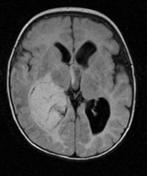
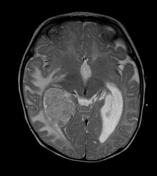
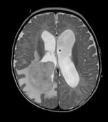
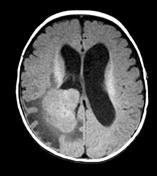
Choroid plexus papilloma

Download
Info

Right lateral ventricular mass centered on the choroid plexus and display iso intense signal on T1, hyperintense signal on T2 and FLAIR with avid enhancement on post contrast study. There is associated unilateral transependymal CSF permeation.


 Unable to process the form. Check for errors and try again.