
Presentation
Presenting history unavailable.
Note: This case has been tagged as "legacy" as it no longer meets image preparation and/or other case publication guidelines.
From the case:
Limbic encephalitis


Download
Info
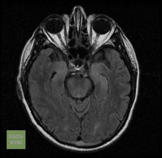
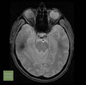
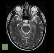
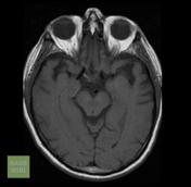
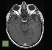
MRI demonstrates bilateral increased signal in the mesial temporal lobes.
Case Discussion
MRI demonstrates bilateral increased signal in the mesial temporal lobes, which in this case was consistent with limbic encephalitis. Unfortunately, clinical details are not available.
This case was donated to Radiopaedia.org by Radswiki.net.


 Unable to process the form. Check for errors and try again.