
Presentation
Right hip pain
Patient Data
Age: Adult
Gender: Male
Note: This case has been tagged as "legacy" as it no longer meets image preparation and/or other case publication guidelines.
From the case:
Transient osteoporosis of the hip
Download
Info

Slight increased density of right femoral head and neck.
Download
Info

Increased uptake of right femur head and neck regions.
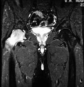
From the case:
Transient osteoporosis of the hip

Download
Info
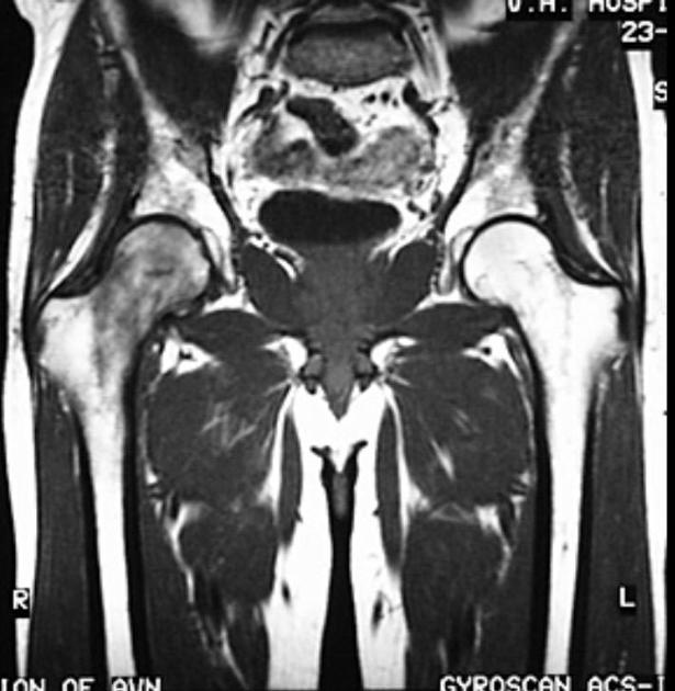
Edema of right femoral head and neck regions.
Case Discussion
Radiograph, bone scan, and MRI images of a patient with right sided transient osteoporosis of the hip.
These images are from Dr. John Hunter's amazing MSK collection. Dr. John Hunter is a professor in the department of radiology (musculoskeletal section) at UC Davis School of Medicine.
This case was donated to Radiopaedia.org by Radswiki.net.


 Unable to process the form. Check for errors and try again.