
Presentation
A 25-years-old man with a history of progressive headaches.
Patient Data
Age: 25 years
Gender: Male


Download
Info
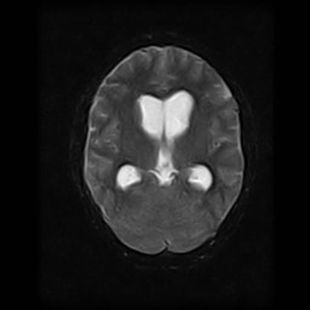
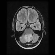
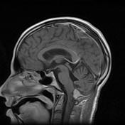
There is a mid-line solid cerebellar mass compressing the 4th ventricle posteriorly and causing hydrocephalus. The mas show vivid contrast enhancement and small flow voids on T2.

Download
Info
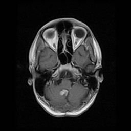
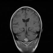
Follow up showing residual/recurrent tumor after resection.
Case Discussion
The diagnosis was confirmed as hemangioblastoma. Eight months after the surgery, a control MRI exams showed signs of relapse.


 Unable to process the form. Check for errors and try again.