
Presentation
Seizure
Patient Data
Age: 50 years
Gender: Male
From the case:
Cavernoma


Download
Info

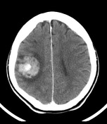
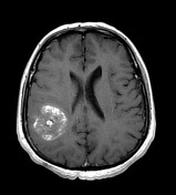
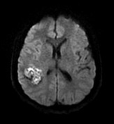
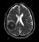
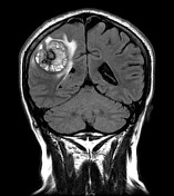
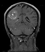
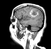
A large right-sided hemorrhagic lesion is present without abnormal enhancement of abnormal vessels. Perilesional edema is noted. Associated mass effect is mild.
From the case:
Cavernoma

Download
Info
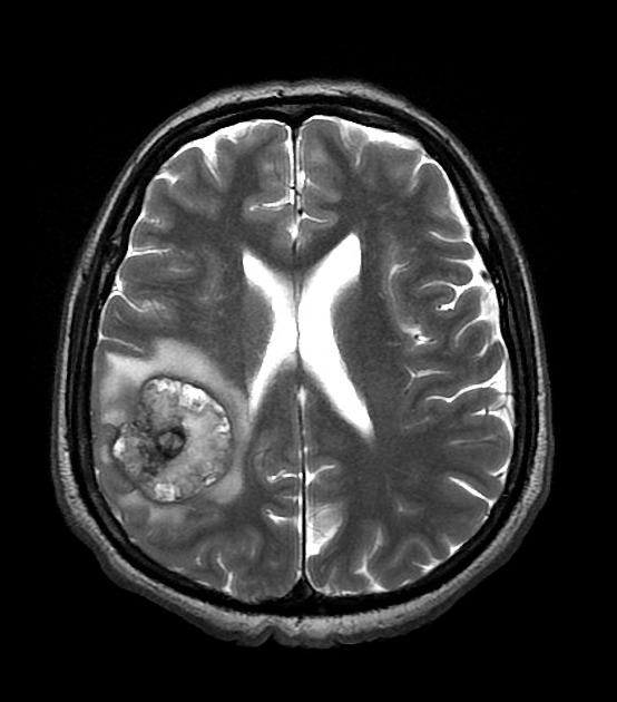
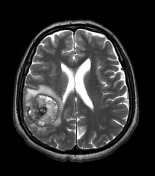
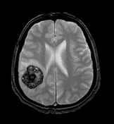
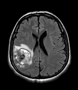
Rounded lesion has extensive hemosiderin staining and surrounding edema. Features are those of a cavernoma.
Case Discussion
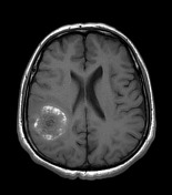
Features are fairly typical of a large cavernous venous abnormality (a.k.a. cavernoma) and edema would be expected to regress. Unfortunately no follow up is available.


 Unable to process the form. Check for errors and try again.