
Presentation
Severe headache, giddiness. On examination the patient had bradycardia and mild papilledema.
Note: This case has been tagged as "legacy" as it no longer meets image preparation and/or other case publication guidelines.


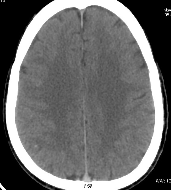
The superior sagittal sinus appears hyperdense on the non-contrast CT images.

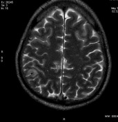
Note loss of flow void in the superior sagittal sinus on T2WI.
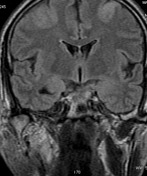
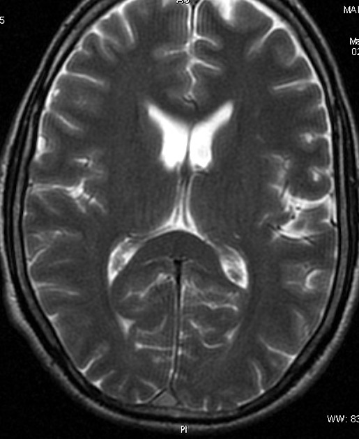
Abnormal hyperintense cortical /subcortical areas on FLAIR images in bilateral high frontal & parietal lobes.
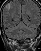
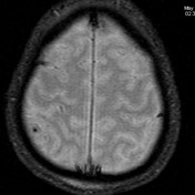
GRE images reveal the 'cord' sign with blooming of the thrombosed superior sagittal sinus (SSS) & cortical veins.
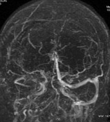
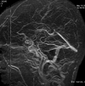
MRV confirms the findings of extensive dural venous sinus thrombosis
Case Discussion
The superior sagittal sinus & a superficial right parietal cortical vein appear hyperdense on noncontrast CT images. Note loss of flow void in the superior sagittal sinus on T2WI. Abnormal hyperintense cortical and subcortical areas on FLAIR images in bilateral high frontal & parietal lobes are also present.
Features are consistent with a dural venous sinus thrombosis.


 Unable to process the form. Check for errors and try again.