
Presentation
Underwent MRI for visual problems. MRI did not reveal parenchymal abnormalities.
Patient Data
Age: 55 years
Gender: Male
Note: This case has been tagged as "legacy" as it no longer meets image preparation and/or other case publication guidelines.
From the case:
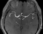
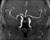
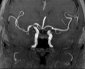
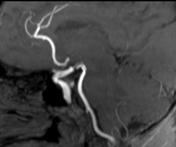
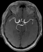
Anterior cerebral arterial variations

Download
Info
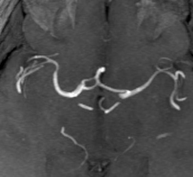
MRI angiography reveals:
- duplicated/fenestrated A1 segment on the right side.
- common A2 trunk which divides into two A3 segments.
- this is different from Azygous ACA - as there is no common trunk for the entire tree (according to Lasjunias-Bernstein classification)
- no ACom
Case Discussion
This case shows two recognized arterial variations of the anterior cerebral artery, fenestrated A1 segment and common trunk of A2 segment, incidentally detected on MR angiography.


 Unable to process the form. Check for errors and try again.