
Presentation
Fall with head trauma.
Patient Data
Age: 90 years
Gender: Female


Download
Info

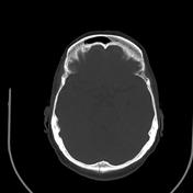
CT shows subarachnoid hemorrhage that extends into the brain parenchyma on the floor of the anterior cranial fossa. Fracture of the occipital bone without bone misalignments. Coronal reformatting CT shows blood in the tent of the cerebellum and within the lateral ventricles.
Download
Info
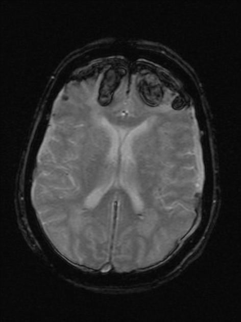
Selected images of MRI study show bilateral frontal contusions and subarachnoid hemorrhage.
Case Discussion
Cerebral hemorrhagic contusion is a type of intracerebral hemorrhage and is common in the setting of significant head injury. This patient probably had a coup-contrecoup mechanism of injury.


 Unable to process the form. Check for errors and try again.