
Presentation
Sudden onset of headache and decreased conscious state.
Patient Data
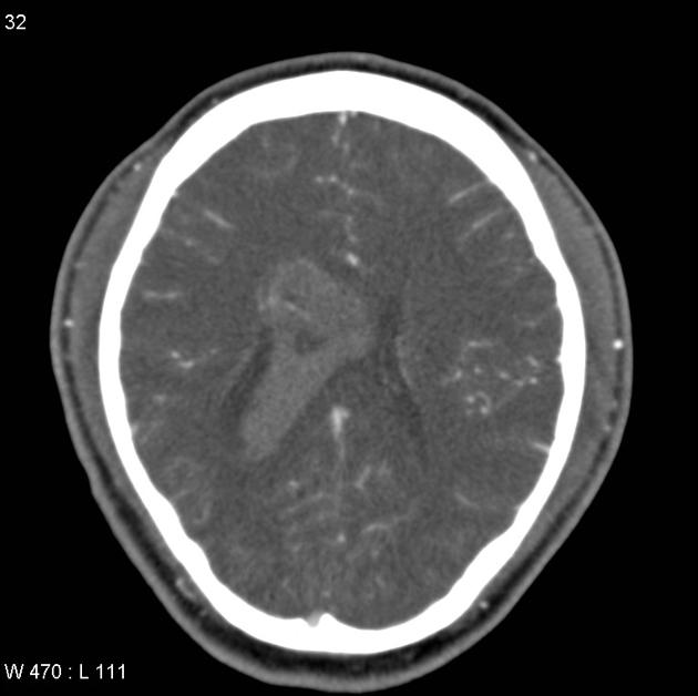
CTA performed for investigation of a non-contrast CT abnormality (not shown) demonstrates florid intraventricular hemorrhage with the impression of a mass in the body of the right lateral ventricle. Hydrocephalus is present with dilatation of the temporal horns of the lateral ventricles.


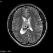
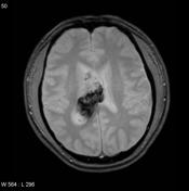
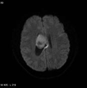

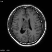
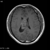
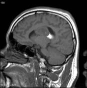
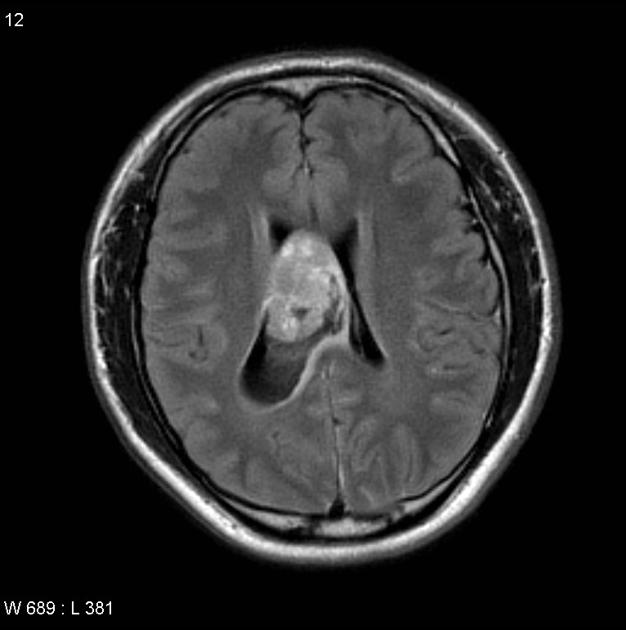
MRI confirms the presence of mass which has a heterogeneous appearance with multiple cysts embedded in a mass which is of increased signal on T2 weighted sequences. Enhancement is present but not prominent and also heterogeneous.
Features suggest a central neurocytoma complicated by hemorrhage, a rare, but recognized complication 1.
Case Discussion
The patient went on to have a resection.
Histology
The sections show multiple fragments of a tumor which shows areas of necrosis and foci of microcalcification. The tumor consists of sheets of regular rounded cells with round nuclei. Chromatin is speckled or vesicular and small nucleoli are often seen. In places, the nuclei surround small rounded eosinophilic areas resembling neuropil. There is also a minor pattern of perivascular pseudorosetting. The mitotic count is elevated at up to 5/10 hpf. There is focal microvascular proliferation.
Immunohistochemical stains show positive staining for synaptophysin and NeuN. The Ki67 stain shows areas of the tumor with up to 15% of nuclei staining.
The features are those of a central neurocytoma. However, the increased proliferative activity, with microvascular proliferation and areas of necrosis suggest that the lesion is best regarded as an atypical neurocytoma. A higher chance of recurrence could be anticipated.


 Unable to process the form. Check for errors and try again.