
Presentation
This young man recalled a history of trauma. He presented with pain and swelling in the left lower leg.
Patient Data
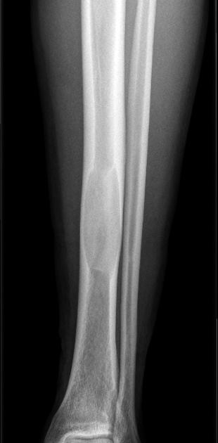
A lytic lesion is demonstrated in the left lower tibial diaphysis with ground-glass appearance, endosteal thinning of the cortex, and some ballooning of the shaft. No periosteal reaction is seen.
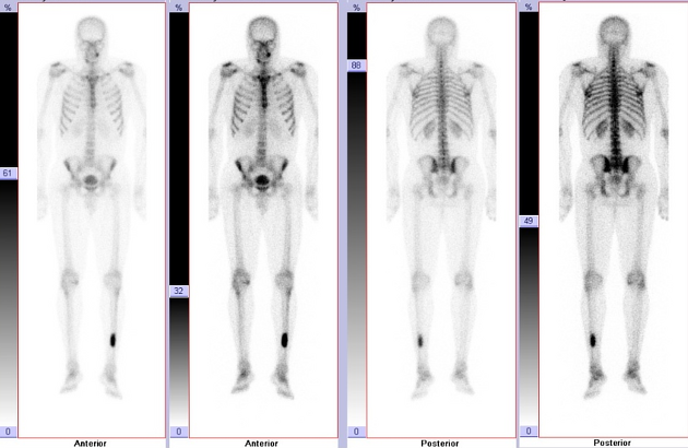
Three phase dynamic bone scintigram (only delayed images shown here) demonstrates a hot spot in the left tibia caused by increased uptake of the radioisotope tracer technetium-99m methylene diphosphonate (99m Tc MDP). The increased uptake is already visible in the early phase.
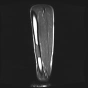

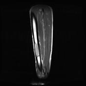

Magnetic resonance imaging (MRI) shows typical features with a low-to-intermediate signal intensity equal to that of muscle on T1-weighted images in the tibial shaft, with endosteal thinning of the cortex and no periosteal or surrounding soft-tissue changes. T2-weighted images also show low signal intensity owing to the high content of collagen and bone. A mild and homogeneous enhancement pattern of the lesion is seen after gadolinium.
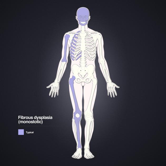
Distribution of monostotic fibrous dysplasia. Layout and distribution: Frank Gaillard 2012, Line drawing of skeleton: Patrick Lynch 2006, Creative Common NC-SA-BY
Case Discussion
Fibrous dysplasia is a skeletal developmental anomaly of the bone-forming mesenchyme that manifests as a defect in osteoblastic differentiation and maturation. Virtually any bone in the body can be affected. It is a nonhereditary disorder of unknown cause.
In this case the diagnosis is straightforward as typical features are present. X-ray is still the examination of first choice. For postoperative follow-up, gadolinium-enhanced MRI can be useful in demonstrating the proliferation of fibrocellular tissue.


 Unable to process the form. Check for errors and try again.