
Presentation
Chronic backaches. HLA-B27 positive.
Patient Data
Age: 40 years
Gender: Male
From the case:
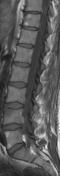
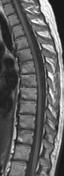
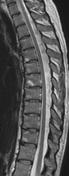
Romanus lesions (MRI)

Download
Info

Active inflammatory spondylitis anterior at L4 and S1 (and posterior at L5). Older, inactive Romanus lesions at L5 and Th 10/11. More metachronous Romanus lesions at Th4-8.
Case Discussion
Spondylitis anterior or Romanus lesions are a typical sign of spondylitis ankylosans (Bechterew disease).


 Unable to process the form. Check for errors and try again.