
Presentation
Progressive right lower neck swelling for 6 months.
Patient Data
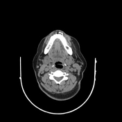

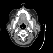
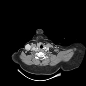
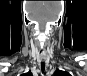

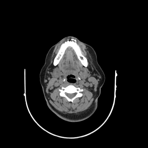
A well-defined oval-shaped lesion is seen in the right lateral lower neck region, measuring about 2.7x2.4 x3.7 cm. The lesion shows gradual enhancement with venous fill-in. It shows linear non-enhancing posterior eccentric filling defect (mostly representing posterior wall thrombus). It is seen connected to the right external jugular vein.
Case Discussion
External jugular vein pseudo-aneurysm is rare condition often caused by trauma.
Doppler US and post contrast CT are the best diagnostic tools for assessment.
Complications of pseudoaneurym include: thrombosis, aneurysm, rupture and thrombo-phlebitis.
Differential diagnosis includes: vascular malformation, abscess, nodal enlargement, lymphocele and schwannoma.


 Unable to process the form. Check for errors and try again.