
Presentation
A lactating female complaining of a palpable left breast lump. No associated pain or signs of inflammation.
Patient Data

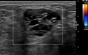
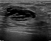
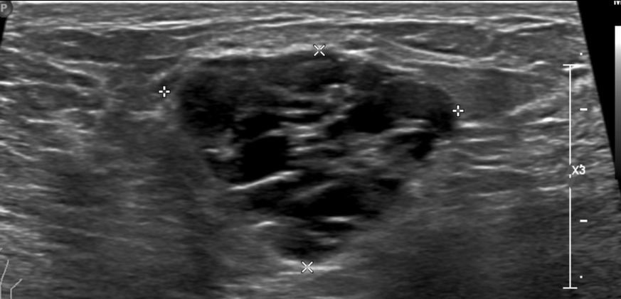
High resolution ultrasound of the left breast shows a well defined micro lobulated predominantly cystic lesion showing clustered microcysts with non-mass lesion located in the left inner quadrant. It measures 14 x 23 x 29 mm.
All cysts appear clear with no floating echoes or calcifications.
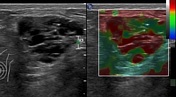
On color Doppler, the lesion showed no definite internal vascularity.
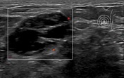
On elastography, it shows no definite signs of stiffness and gives BGR (blue-green-red) signal.
Case Discussion
Clustered microcysts are not that common, account for about 6 % of breast lesions, and are almost benign. Its origin is the terminal lobular unit that shows cystic dilatation.
The lesion term clustered microcysts should fulfill a few criteria as the cysts should be tiny, 2-3 mm with intervening septae less than 0.5 mm and showing no definite solid components.
This case was proved by histopathology to be lactational adenosis/lactational adenoma associated with fibrocystic changes and apocrine metaplasia.
BI-RADS 2: benign lesion (reporting system of UK National Health Service Screening Programme)
When should we recommend a biopsy for microcystic clusters?
In post menopausal women and if there are any suspicious signs as solid components, hyper vascular thickened internal septae. Otherwise, annual ultrasound follow is recommended.


 Unable to process the form. Check for errors and try again.