
Presentation
Menometrorrhagea.
Patient Data
Age: 35 years
Gender: Female
From the case:
Prolapsed endometrial polyp


Download
Info
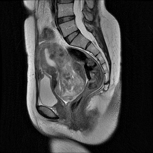
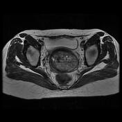
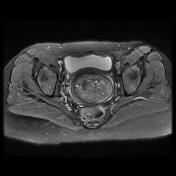
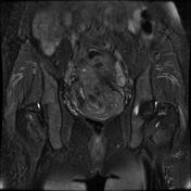
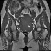
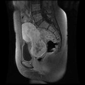
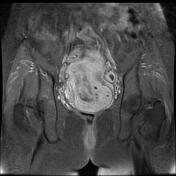
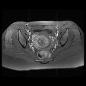
Large pedunculated intracavitary mass arising from the uterine posterior wall, filling the uterine cavity and prolapsing into the vagina through a distended endocervical canal.
The postcontrast sequences show a heterogeneous enhancement.
Few intramural myomas with maximum diameters up to 10 mm are seen (Figo IV).
Intramural submucosal myoma measuring about 20 mm along with uterus left anterolateral body (Figo II).
No pelvic lymphadenopathy is seen.
Case Discussion
MRI features are most consistent with a prolapsed endometrial polyp.


 Unable to process the form. Check for errors and try again.