
Presentation
Chronic upper abdominal pain. History of surgical excision of hepatic hydatid cyst.
Patient Data
Age: 35 years
Gender: Female
From the case:
Hepatic hydatid cyst


Download
Info
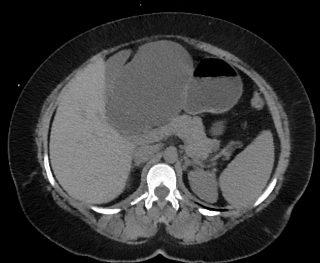
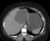
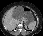
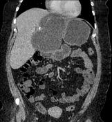
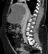
A large hypoattenuating non-enhancing cystic lesion is seen occupying most of the left liver lobe with large exophytic component, multiple fine internal septations, peripheral coarse calcifications as well as septal calcifications. The mass is seen encroaching upon the porta hepatis, compressing the CHD & CBD with subsequent minimal dilatation of the IHBRs. This mass shows the following relations:
- posteriorly & inferiorly seen contacting the gastric pylorus, the first part of the duodenum, the main portal vein, and the anterior aspect of the pancreatic head & neck.
- left laterally seen contacting the gastric lesser curvature.
- anteriorly & right laterally contacting the gall bladder.
Case Discussion
CT findings are highly suggestive of hepatic hydatid cyst.
This patient had a past history of two times surgical excision of similar hepatic cysts which were pathologically proved to be hydatid cysts.


 Unable to process the form. Check for errors and try again.